

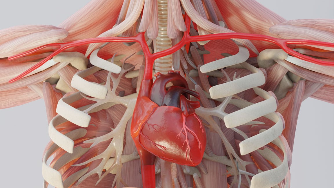

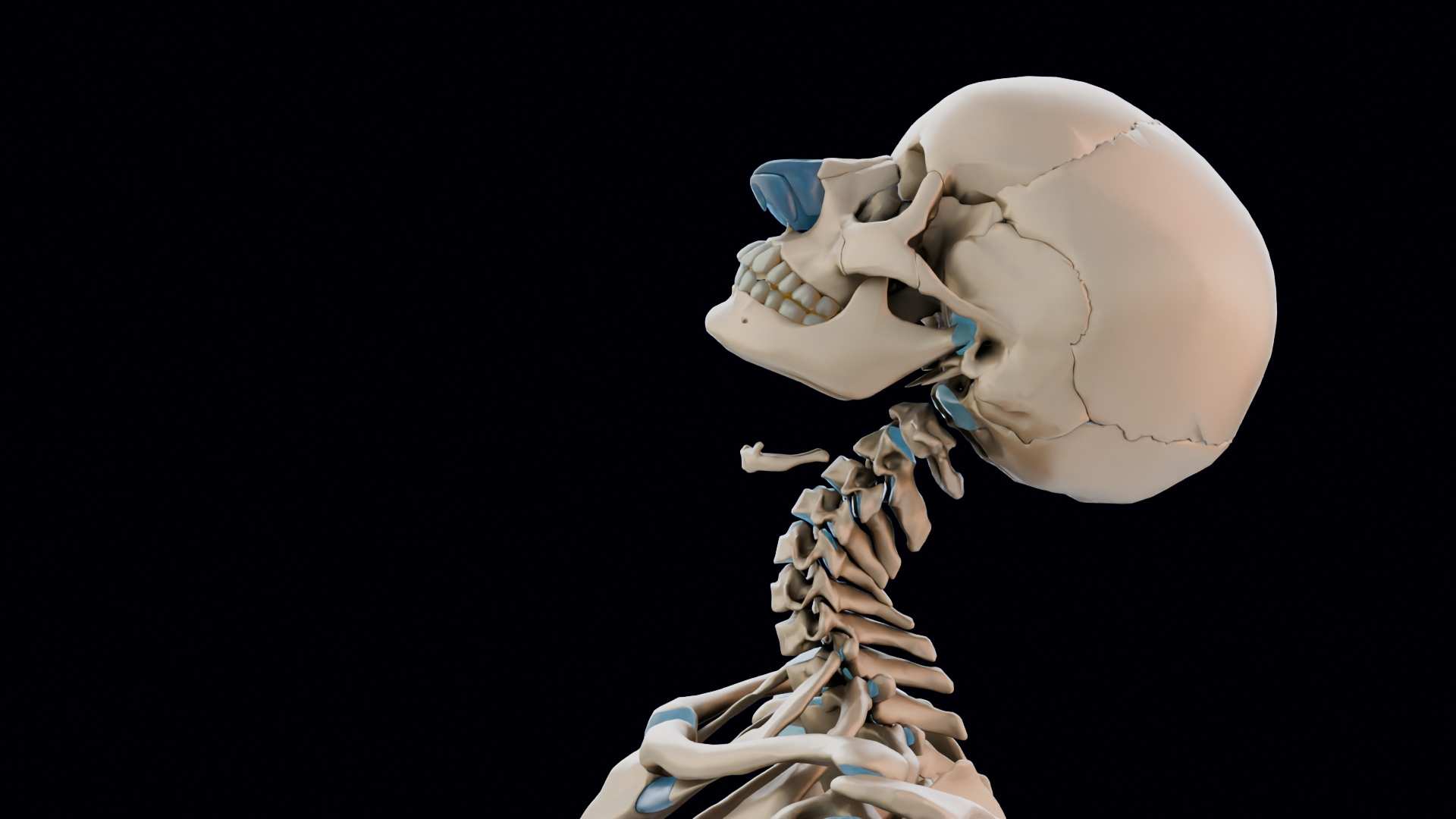
Spinal fusion is often the biggest number in the case, and the hardest to explain.
The jury hears “C5–C6 herniation” and “anterior cervical discectomy and fusion” and pictures very little. We show the injury, the compressed nerve and every step of the surgery, built from your client’s own MRI and operative report.
Part 1 · The injury
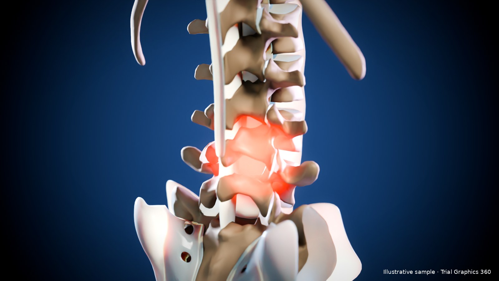
From the mechanism to the herniated disc
- 1Mechanism. The seat-back load, the S-curve of the neck, extension and rebound, or the fall or lift that caused it.
- 2Disc herniation. The annular tear and disc material pushing into the canal or foramen, from your client’s MRI.
- 3Nerve compression. The root or cord it presses on, so the jury connects the image to the symptoms.
- 4Pre-existing vs new. Earlier and later imaging side by side, so your expert can show what changed.
Part 2 · The surgery
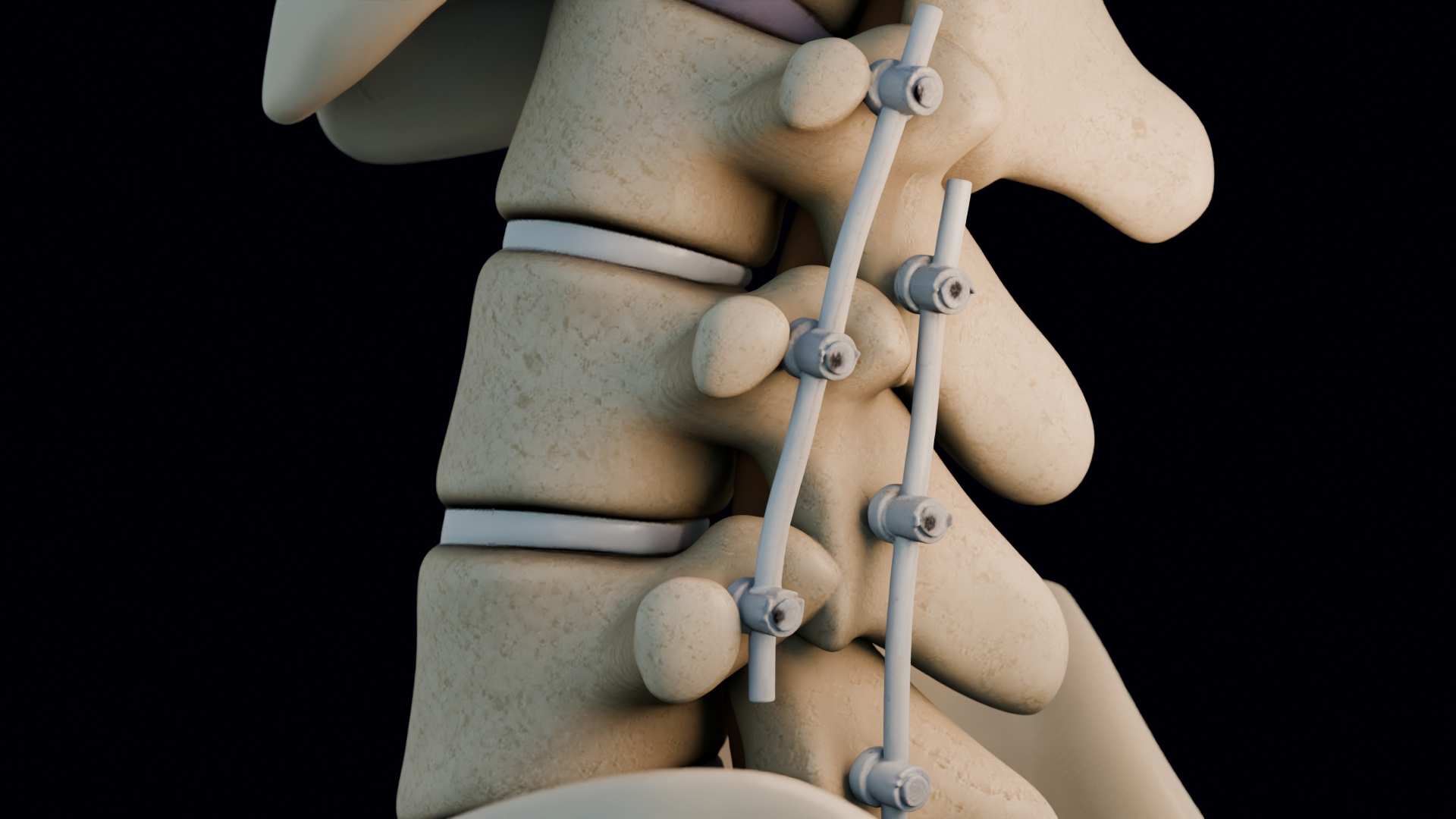
Every step of the ACDF or fusion
- 1The approach. Anterior for ACDF; posterior, anterior or lateral for lumbar fusion, as the op note records.
- 2Decompression. The disc removed and the nerve root freed.
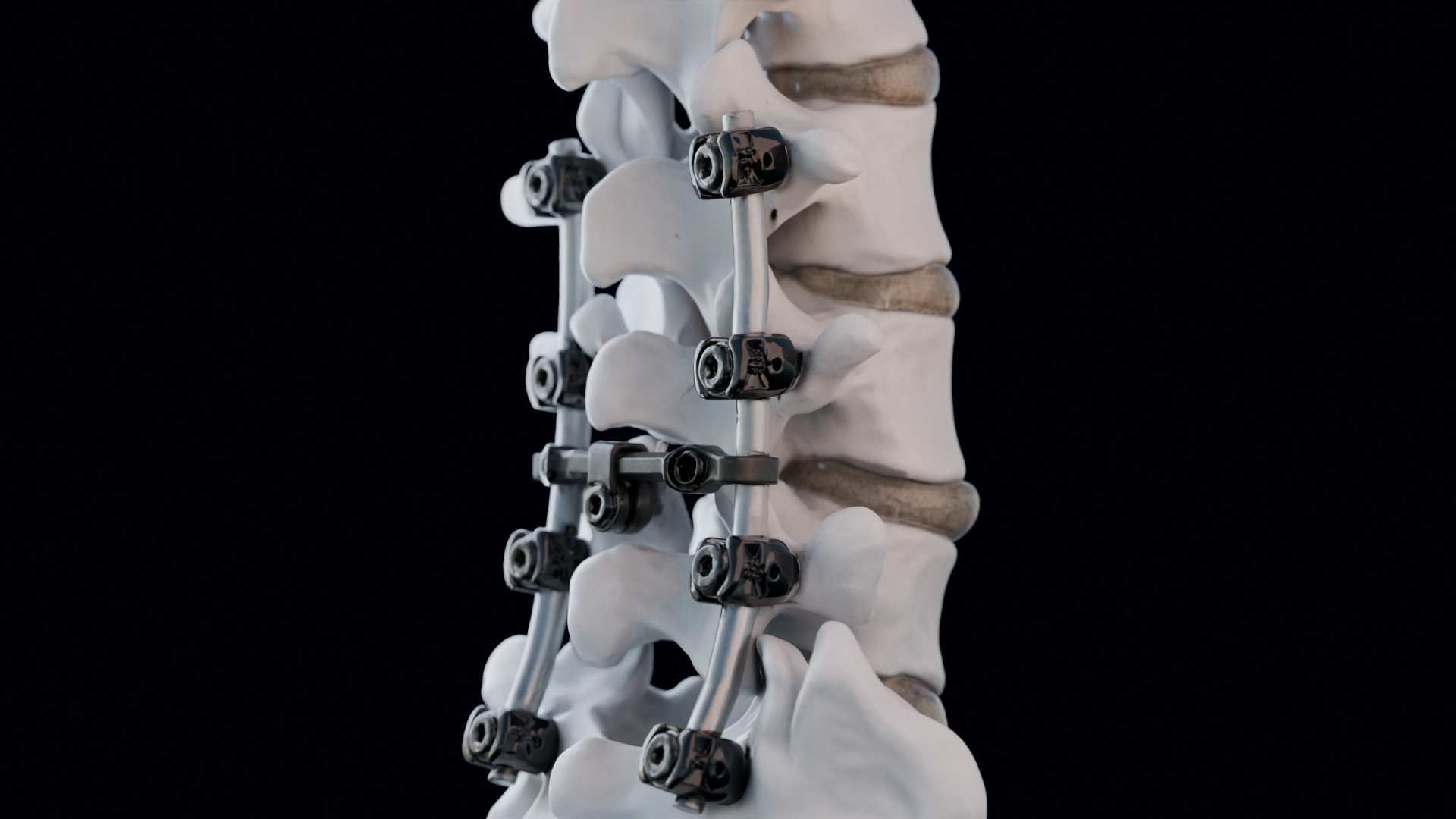
- 3Fixation. Cage or graft, plate, pedicle screws and rods, sized from the implant log.
- 4Future surgery. Adjacent segment disease and further surgery your expert supports, labelled as future treatment.
Crash, injury and surgery in one sequence, or separate clips so each witness can use their part.
See it in motion
From the crash to the herniated disc
 Whiplash to Disc Herniation: A 30 km/h Rear-End CrashA low-speed rear-end impact, the occupant’s neck motion, and the disc herniation it caused, in one continuous sequence.
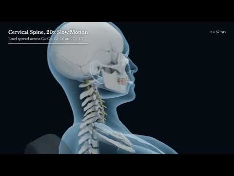 One Crash, Three Herniated Discs: A 50 km/h Rear-End CollisionHow a single rear-end collision can injure several levels of the spine, shown from impact to MRI findings.
Accuracy
Built from your client’s own imaging
The right level and side
The herniation is shown at the level and on the side your client’s MRI shows, not a textbook default.
Sized from the records
Cages, plates and screws are sized from the implant log and placed to match the post-op films.
Future care labelled
Anything not yet done is shown clearly as recommended future treatment.
How it works
From Records To Courtroom, On Your Deadline
- Step 01
Send the records
MRI and CT, op report, implant log, crash findings and your dates.
- Step 02 · 12 hours
Visual plan and fixed fee
A shot-by-shot plan and a fixed price, before any work starts.
- Step 03
Build and expert review
Your expert checks each stage before anything is disclosed.
- Step 04
Delivered for mediation or trial
Video, stills and exhibit boards, with source notes for admissibility.
What We Need To Start
- MRI and CT (DICOM if possible)
- Radiology reports
- Operative report and implant log
- Crash or incident findings
- Your expert’s report and your dates
Send the records. Get a visual plan and fixed-fee quote within 12 business hours.
Free, with no obligation. Tell us your date first and we’ll tell you honestly what can be built in time.

Clinician-directed
Every animation is directed by Dr Kevin Ho, who reads the op note the way your expert does.
Our medical work has been published alongside Harvard Medical School and Sanofi.
Common Questions
Can you show the crash and the surgery in one animation?
Yes. We can carry the collision through to the injury and the surgery in one sequence, or deliver separate clips so each witness can use their part.
My client hasn’t had surgery yet. Can you show future surgery?
Yes, as long as a treating surgeon or life-care planner supports it. We label it clearly as recommended future treatment.
Can it be a still exhibit instead of a full animation?
Yes. Any frame can be exported as a still, and a short set of stills is often enough for mediation.
How fast can you deliver?
You get a free visual plan and fixed-fee quote within 12 business hours. Delivery depends on scope and your mediation or trial date, and we’ll tell you up front if a deadline is tight.