


Spine cases don't lose because the injury isn't real. They lose because the jury can't feel pain — and a static film doesn't show them why your client does.
Surgical Hardware & Fusion Reconstructions
Show the Jury Not Just That Hardware Is There — But What It Did, What It Changed, and What It Failed to Fix.
Post-operative spine cases require more than showing a screw on an X-ray. We reconstruct the full surgical intervention — the anatomy before, the procedure itself, and the mechanical reality after — so the jury understands the permanence of the alteration and the consequences your client now lives with. Instrumentation Visualization Patient-specific 3D reconstruction of pedicle screws, rods, interbody cages, and plates as they actually sit within your client's post-operative anatomy — derived directly from DICOM imaging. Malposition, proximity to nerve roots, and hardware-to-bone relationships are all rendered with clinical precision. Biomechanical Impact We animate how a fusion alters the natural load distribution of the spine — showing how immobilized segments transfer stress to adjacent levels, the biomechanical basis for Adjacent Segment Disease, and why hardware failure or revision surgery is a foreseeable consequence rather than an unrelated event. Hardware Failure In product liability and surgical negligence cases, we visualize the mechanical fatigue, fracture, or migration of orthopedic implants — using post-operative imaging to show exactly where the hardware failed, what it was adjacent to, and what the failure caused structurally and neurologically.
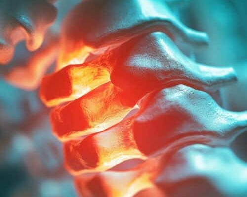
Nerve Root Compression & Disc Pathology
Pain Is Invisible. The Mechanism That Causes It Is Not — Once You Can See the Nerve.
Radiculopathy, myelopathy, and permanent neurological deficits are the most contested elements of spine cases. The defense argues the symptoms are exaggerated. We show the jury exactly what is compressing the nerve, by how much, and which symptoms that compression anatomically explains — so your expert's opinion becomes something the jury can see for themselves. Compression Visualization We render the exact point where a herniated disc, osteophyte, or displaced fracture fragment physically impinges on the thecal sac or exiting nerve root — derived from your client's MRI data. The jury sees the compression in three dimensions, not just hears about it in testimony. Dermatome Mapping A 3D dermatome map linking the spinal level of injury to the exact areas of your client's body affected — showing how a compression at C5-C6 explains the arm weakness, how an L4-L5 herniation explains the leg pain, and why the distribution of symptoms is anatomically consistent with the structural injury documented in the imaging. Pre-existing vs. Traumatic Pathology Side-by-side comparison of imaging taken before and after the incident — isolating the new traumatic pathology from any pre-existing degenerative changes. The defense argument that the injuries were pre-existing is directly rebutted by the imaging chronology itself, rendered in three dimensions.
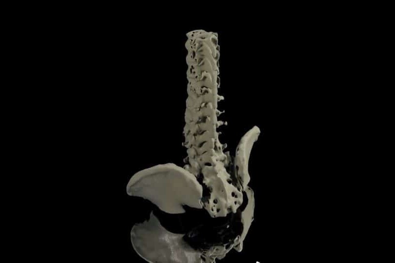
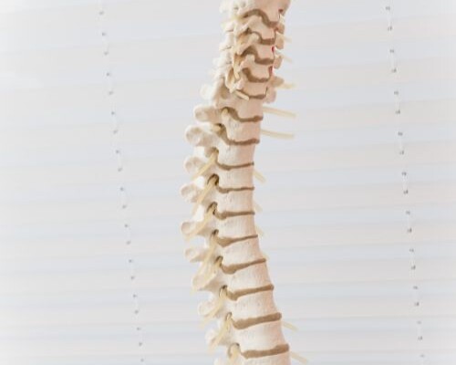
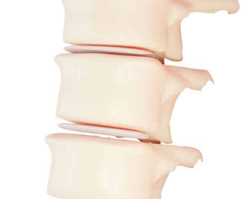
Complex Fractures & Pelvic Trauma
When the Bone Is Shattered Into Fragments — We Reassemble It in 3D So the Jury Sees Every Piece.
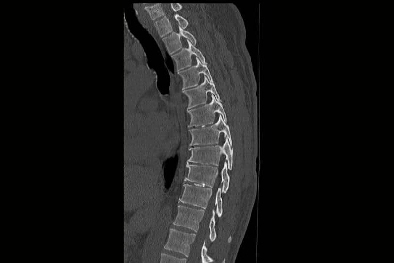
Comminuted fractures, burst fractures, and pelvic ring disruptions are among the most visually complex injuries to present to a lay jury. A 2D film shows gray shadows. We use Hounsfield Unit thresholding to isolate every fragment from the raw DICOM data and render the full fracture pattern in three dimensions — so the severity of the impact, the instability of the injury, and the permanence of the consequences are all immediately visible. Comminuted Fracture Reconstruction Every bone fragment is isolated using Hounsfield Unit thresholding applied directly to your client's CT data — then rendered in 3D to show the full fracture pattern, fragment displacement, and proximity to vascular structures, nerve roots, and the spinal canal. The severity of the impact becomes spatially undeniable. Pelvic & Long-Bone Stability Disruption of the pelvic ring, intra-articular joint involvement, and long-bone fracture patterns are reconstructed in three dimensions — showing the instability of the injury, the surgical complexity of the repair, and the long-term risk of post-traumatic arthritis and avascular necrosis that your damages expert will testify to. Surgical Repair & Residual Deficit We animate the surgical repair of complex fractures alongside the post-operative anatomy — showing what was fixed, what could not be fully restored, and what structural compromise remains. The gap between the pre-injury anatomy and the post-surgical reality becomes the visual foundation for your damages argument.
Every Animation Is Built From Your Actual Case Records — Not Generic Templates.
Unlike generic animations, our demonstratives are reconstructed from the actual clinical record.
Operative Reports
Step-by-step surgical reconstructions from case documentation.
CT & MRI Imaging
Accurate, DICOM-derived 3D reconstructions of patient anatomy and surgical outcomes.
Expert Testimony
Every animation is developed in collaboration with your retained experts and evolves alongside their opinions.
Medical Literature
Science-backed visualizations grounded in peer-reviewed evidence.
Dedicated Medical Experts For Every Case
Dr. Kevin Ho and his own team of medical experts collaborates directly with your experts for scientifically defensible, strategically aligned medical animations.
Technical Alignment
Every visual element is reviewed against expert reports, deposition testimony, imaging findings, and evolving case facts.
Built on Defensible Science
Animations are built using transparent reconstruction techniques that experts can confidently explain and defend.
Litigation-Focused Collaboration
We help translate highly technical medical concepts into clear visual narratives that resonate with judges, mediators, and juries.
We Stay With Your Case From First Filing to Final Verdict.
Medical negligence cases take years. Expert opinions evolve. New imaging comes in. The case theory shifts. We stay engaged for the full life of the case — refining, updating, and adapting your visual evidence at every stage.
Evolving Forensic Models
As new records, imaging, or expert findings emerge, we update the master 3D models to reflect the most current picture of the injury. Your animation never becomes a liability because it was built too early.
Ongoing Litigation & Deposition Support
We build animations to support long-term litigation — not a single courtroom moment. Demonstratives are adapted through discovery, expert depositions, mediation, and trial, keeping testimony consistent from beginning to end.
The Scientific Credibility Your Opposing Experts Will Recognize
Our work is published by the same institutions cited in medical literature and expert reports. That rigor is what makes our animations defensible — not just persuasive.


Why Top Law Firms Partner with Trial Graphics 360
Unmatched experience and clarity for your case's legal animations.
$100M+
Increased settlement value in high-stakes litigation.
Medically-Trained Animators
Board-certified specialists and former UBC professors ensuring medical accuracy for complex litigation.
Scientifically Published
Harvard, Stanford, and pharma-published expertise giving your case a distinct litigation edge.
Long Term Case Support
End-to-end case support, refining visuals as evidence evolves.
Common Questions About Colorized Diagnostic Films
It is the most common defense argument in spine litigation and we address it directly in every reconstruction. We build side-by-side comparisons of pre-incident and post-incident imaging — isolating the new traumatic pathology from any documented degenerative changes and showing the jury exactly what the incident caused that was not there before. The chronological imaging record rebuts the defense argument visually, not just verbally.
Yes. Through dermatome mapping, range-of-motion animations, and functional anatomy visualizations, we connect the structural injury to the specific symptoms and limitations your client experiences — showing the jury the anatomical basis for every deficit your client and their treating physicians have described.
Pre- and post-operative CT and MRI DICOM files are ideal. For hardware cases, post-operative CT provides the most accurate implant positioning data. We work with whatever imaging is available and advise on whether additional imaging would strengthen the reconstruction before expert depositions.
Yes. Our methodology is fully documented — Hounsfield Unit thresholding parameters, DICOM source data, hardware positioning derivation, and biomechanical literature citations are all recorded. Dr. Ho can provide a complete methodology report and your expert can defend every frame from the stand.
Most spine and orthopedic animations are completed in 4–8 weeks depending on the complexity of the surgical history and the volume of imaging data. Rush delivery is available — send us your mediation or trial date and we will confirm availability the same day.