

Every colorized film is reviewed by a clinician — not a graphic designer. The pathology we highlight matches your expert's findings exactly, so nothing in the image contradicts testimony.

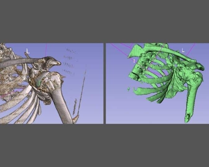
Orthopedic & Fracture Enhancement
Make Fractures and Hardware Failures Impossible to Dispute.
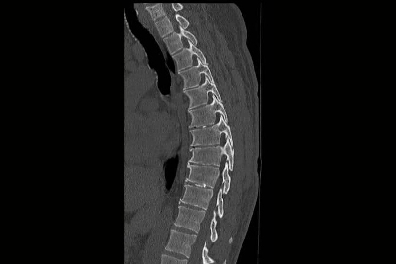
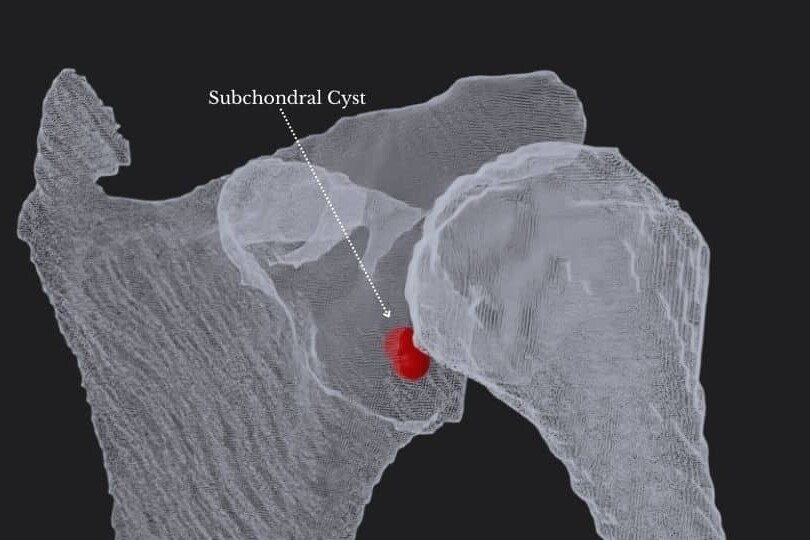
Hairline fractures and complex shatter patterns disappear in standard grayscale film. We apply clinical colorization so the jury sees the structural failure your expert has been describing — clearly, immediately, and without needing a radiology degree. Fracture Visualization High-contrast color applied to hairline fractures, compression fractures, and complex shatter patterns that are routinely missed by untrained eyes on standard film. Your expert points to it. The jury sees it. Hardware Verification Distinct color-coding of surgical screws, plates, rods, and cages showing their exact orientation relative to bone — making malposition, loosening, or failure visible without expert narration alone. Density Mapping Color gradients applied to areas of decreased bone density, non-union at a surgical site, or avascular necrosis — translating Hounsfield Unit data into a visual the jury can interpret without technical guidance.
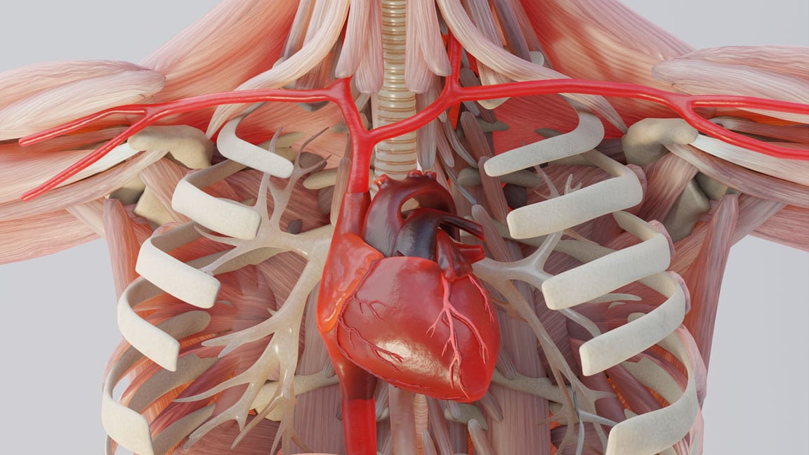
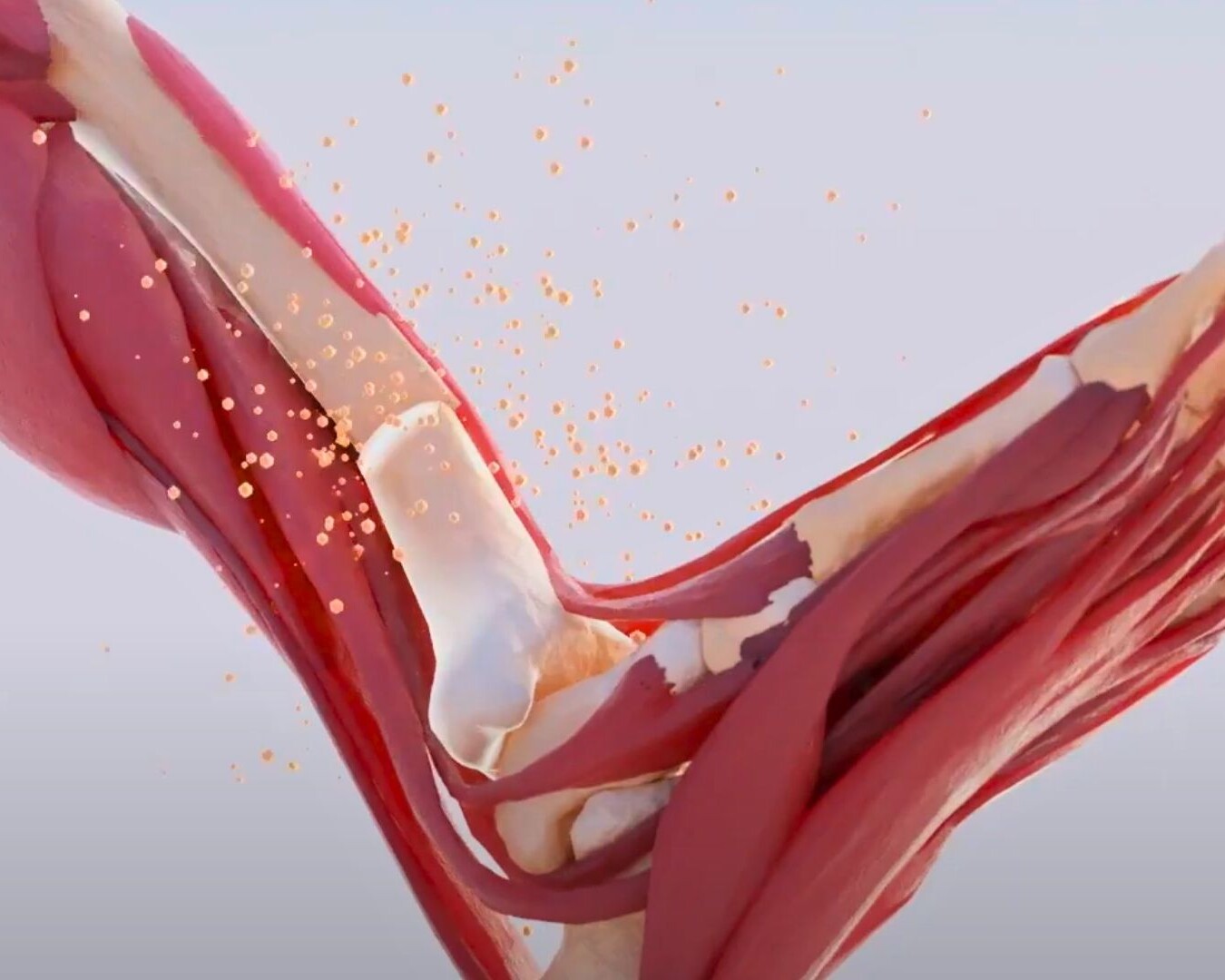
Soft Tissue & Neurological Pathology
The Injuries That Don't Show Up on a Standard Film — Until We Enhance Them.
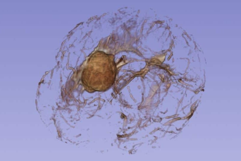
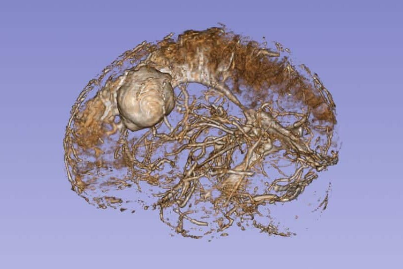
Soft tissue injuries are the hardest to prove because they're the hardest to see. Disc herniations, nerve compression, brain bleeds, and ligament tears are real, documented, and devastating — but to a juror, they look like slightly different shades of gray. We change that. Disc & Nerve Compression We colorize the exact point where a herniated disc impinges on a nerve root in a spinal MRI — so the jury sees the compression, not just hears about it. The relationship between the disc and the nerve becomes spatially clear. Hematoma & Edema Distinct color borders applied to brain bleeds, subdural hematomas, and edema — defining their size, location, and proximity to critical structures. The severity of the injury becomes visually undeniable. Ligament & Tendon Tears The subtle signal changes in an MRI that indicate a full or partial tear are enhanced and highlighted so the jury understands what your expert means when they say the structure is ruptured — not just strained.
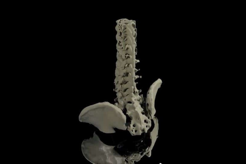
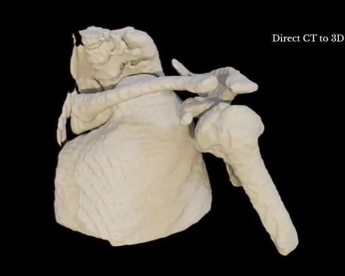
3D Volumetric Overlays
Give a 2D Film a 3D Perspective — So the Jury Understands Where the Injury Actually Is.
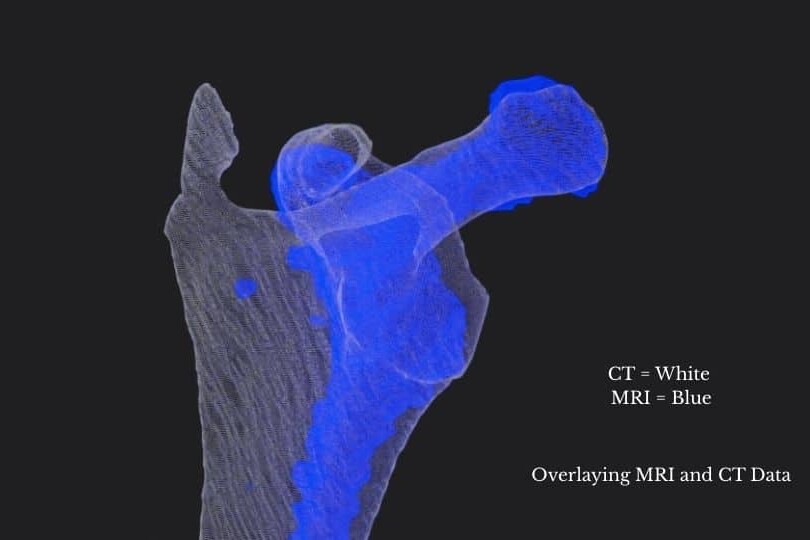
A flat X-ray or MRI slice gives no spatial context. Jurors can't tell depth, orientation, or how one structure relates to another. We add depth, dimension, and anatomical reference so your client's imaging tells a complete story. Depth & Spatial Orientation 3D shadowing and perspective applied to flat X-rays so the jury understands where the injury sits in the body — not just that it exists. Spatial context is what makes causation believable. Anatomical Mapping A colorized 3D anatomical model overlaid on the 2D scan — giving the jury a visual reference point for every structure your expert identifies. Think of it as a legend for the film. Side-by-Side Comparison The original grayscale film paired with the enhanced version in a single exhibit. The before-and-after format is the most effective courtroom format for this type of evidence — opposing counsel sees the same thing the jury sees.
Every Animation Is Built From Your Actual Case Records — Not Generic Templates.
Unlike generic animations, our demonstratives are reconstructed from the actual clinical record.
Operative Reports
Step-by-step surgical reconstructions from case documentation.
CT & MRI Imaging
Accurate, DICOM-derived 3D reconstructions of patient anatomy and surgical outcomes.
Expert Testimony
Every animation is developed in collaboration with your retained experts and evolves alongside their opinions.
Medical Literature
Science-backed visualizations grounded in peer-reviewed evidence.
Dedicated Medical Experts For Every Case
Dr. Kevin Ho and his own team of medical experts collaborates directly with your experts for scientifically defensible, strategically aligned medical animations.
Technical Alignment
Every visual element is reviewed against expert reports, deposition testimony, imaging findings, and evolving case facts.
Built on Defensible Science
Animations are built using transparent reconstruction techniques that experts can confidently explain and defend.
Litigation-Focused Collaboration
We help translate highly technical medical concepts into clear visual narratives that resonate with judges, mediators, and juries.
We Stay With Your Case From First Filing to Final Verdict.
Medical negligence cases take years. Expert opinions evolve. New imaging comes in. The case theory shifts. We stay engaged for the full life of the case — refining, updating, and adapting your visual evidence at every stage.
Evolving Forensic Models
As new records, imaging, or expert findings emerge, we update the master 3D models to reflect the most current picture of the injury. Your animation never becomes a liability because it was built too early.
Ongoing Litigation & Deposition Support
We build animations to support long-term litigation — not a single courtroom moment. Demonstratives are adapted through discovery, expert depositions, mediation, and trial, keeping testimony consistent from beginning to end.
The Scientific Credibility Your Opposing Experts Will Recognize
Our work is published by the same institutions cited in medical literature and expert reports. That rigor is what makes our animations defensible — not just persuasive.


Why Top Law Firms Partner with Trial Graphics 360
Unmatched experience and clarity for your case's legal animations.
$100M+
Increased settlement value in high-stakes litigation.
Medically-Trained Animators
Board-certified specialists and former UBC professors ensuring medical accuracy for complex litigation.
Scientifically Published
Harvard, Stanford, and pharma-published expertise giving your case a distinct litigation edge.
Long Term Case Support
End-to-end case support, refining visuals as evidence evolves.
Common Questions About Colorized Diagnostic Films
Yes. Colorized diagnostic films are demonstrative exhibits — they illustrate testimony, not replace it. Admissibility is supported when the enhancement is clinically directed, methodologically transparent, and aligned with your expert’s findings. Our process is built to meet that standard.
It is fundamentally different. We work from raw DICOM data using Hounsfield Unit thresholding — meaning color is applied based on actual tissue density values from the scan, not artistic judgment. Every enhancement decision is made by Dr. Ho and cross-referenced against your expert’s report.
Yes — and mediation is often where they are most effective. When opposing counsel sees the injury clearly for the first time, the conversation about settlement value changes quickly.
We work with all standard DICOM formats including CT, MRI, X-ray, and CBCT. If you have the imaging, we can work with it.
Most colorized film projects are completed in 2–4 weeks depending on volume and complexity. Rush delivery is available — send us your mediation or trial date and we will confirm availability the same day.