


Every reconstruction is built from raw DICOM data using Hounsfield Unit thresholding — not artistic interpretation. The methodology is fully documented and defensible by your expert on the stand.
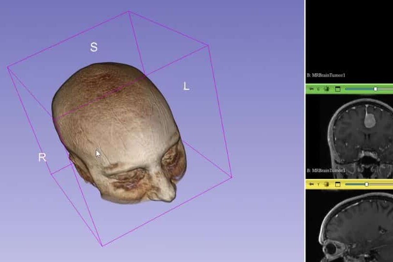
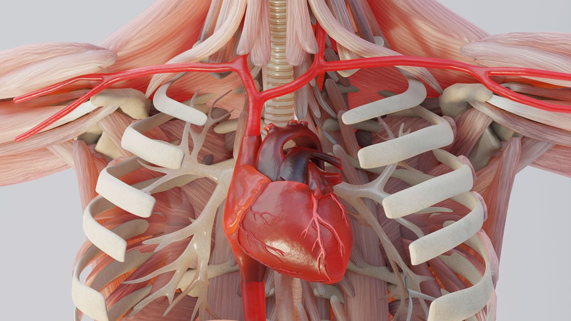
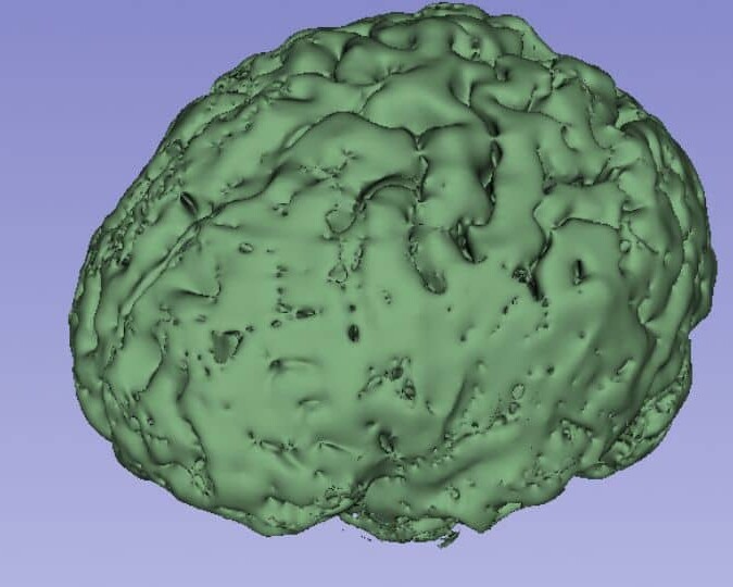
Volumetric & Surface Rendering
Turn Flat Grayscale Slices Into a 3D Model the Jury Can Actually See Inside.
We use advanced volumetric rendering and 3D Slicer technology to convert raw DICOM imaging into navigable three-dimensional anatomy. The result is a forensic model your expert can rotate, zoom, and dissect in front of the jury — built entirely from your client's own scan data. Data Integrity Direct conversion from DICOM metadata into high-resolution 3D geometry — no guesswork, no artistic interpolation. The geometry is derived from actual pixel density values in the original imaging data, making every structural detail defensible against a radiology expert challenge. Tissue Segmentation Bone, soft tissue, vascular structures, and surgical hardware are individually segmented and color-coded — so your expert can isolate and highlight the exact injured structure without surrounding anatomy obscuring the view. Spatial Relationship The precise proximity of hardware to nerves, a tumor to a vessel, or a fracture fragment to the spinal cord is rendered in three-dimensional space — so the jury understands not just that an injury exists, but exactly where it is and what surrounds it.
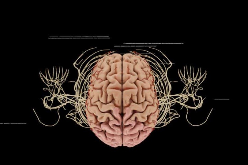
Colorized Diagnostic Enhancement
The Injuries That Disappear on Standard Film — Made Impossible to Miss in 3D.
Grayscale imaging requires a trained eye. We apply clinical colorization and density-based enhancement directly to the 3D model — so the pathology your expert has been describing becomes something the jury can see with no training at all. Clinical Colorization Color is applied based on Hounsfield Unit thresholding — actual tissue density values from the scan — not artistic choice. Fractures, herniations, bleeds, and lesions are highlighted in a way that is both visually clear and scientifically grounded. Pathology Isolation The exact borders of a hematoma, lesion, or infection are defined within the 3D model and rendered in distinct color — so the size, location, and proximity to critical structures are immediately apparent to any decision-maker in the room. Interactive Views Individual tissue layers can be peeled back to reveal deep-seated orthopedic or neurological trauma — giving your expert the ability to show the jury the injury from any angle, at any depth, in real time during testimony.
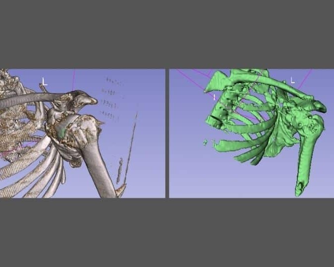
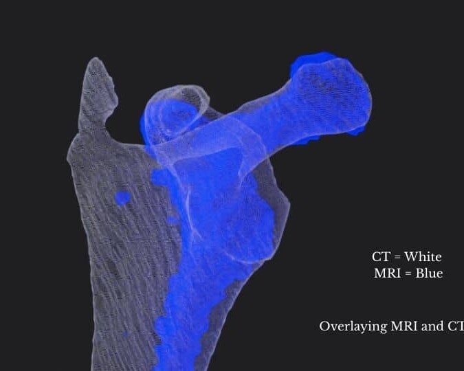
Comparative & Forensic Analysis
Side-by-Side Comparisons That Make the Extent of Damage Undeniable.
The most powerful exhibits in a radiology reconstruction case are direct comparisons — normal anatomy versus your client's anatomy, pre-operative versus post-operative, defendant's imaging interpretation versus your expert's. We build all three. Normal vs. Patient A side-by-side 3D comparison of standard anatomical structure versus your client's actual anatomy — so the deviation from normal is visually self-evident without your expert needing to explain what healthy looks like to a lay jury. Hardware Verification Accurate 1:1 rendering of surgical screws, plates, rods, and implants relative to the surrounding bone and soft tissue — showing malposition, loosening, or proximity to critical structures with the precision of the original DICOM data. Pre- vs. Post-Op Anatomical models rendered from imaging taken before and after a surgical intervention or traumatic event — showing exactly what changed, what was altered, and what the intervention did or failed to correct.
Dedicated Medical Experts For Every Case
Dr. Kevin Ho and his own team of medical experts collaborates directly with your experts for scientifically defensible, strategically aligned medical animations.
Technical Alignment
Every visual element is reviewed against expert reports, deposition testimony, imaging findings, and evolving case facts.
Built on Defensible Science
Animations are built using transparent reconstruction techniques that experts can confidently explain and defend.
Litigation-Focused Collaboration
We help translate highly technical medical concepts into clear visual narratives that resonate with judges, mediators, and juries.
We Stay With Your Case From First Filing to Final Verdict.
Medical negligence cases take years. Expert opinions evolve. New imaging comes in. The case theory shifts. We stay engaged for the full life of the case — refining, updating, and adapting your visual evidence at every stage.
Evolving Forensic Models
As new records, imaging, or expert findings emerge, we update the master 3D models to reflect the most current picture of the injury. Your animation never becomes a liability because it was built too early.
Ongoing Litigation & Deposition Support
We build animations to support long-term litigation — not a single courtroom moment. Demonstratives are adapted through discovery, expert depositions, mediation, and trial, keeping testimony consistent from beginning to end.
The Scientific Credibility Your Opposing Experts Will Recognize
Our work is published by the same institutions cited in medical literature and expert reports. That rigor is what makes our animations defensible — not just persuasive.


Why Top Law Firms Partner with Trial Graphics 360
Unmatched experience and clarity for your case's legal animations.
$100M+
Increased settlement value in high-stakes litigation.
Medically-Trained Animators
Board-certified specialists and former UBC professors ensuring medical accuracy for complex litigation.
Scientifically Published
Harvard, Stanford, and pharma-published expertise giving your case a distinct litigation edge.
Long Term Case Support
End-to-end case support, refining visuals as evidence evolves.
Common Questions
A radiologist reads and reports on imaging for clinical purposes. We convert that same imaging data into a three-dimensional exhibit built for a lay audience — rotating, interactive, and clinically annotated so a jury can understand the injury without a medical degree. The radiologist reads it. We make it visible.
Yes. Because the reconstruction is derived directly from the raw imaging data using documented, transparent methodology — not artistic interpretation — it can be defended as a scientifically accurate representation of the plaintiff’s actual anatomy. Dr. Ho can provide a full methodology report and your expert can defend it from the stand.
We work with all standard DICOM formats including CT, MRI, CBCT, and X-ray. If you have the imaging, we can work with it.
Yes. We maintain the master source files throughout the life of the case. As new imaging is produced — through follow-up care, revision surgery, or independent medical examination — we update the model to reflect the most current anatomical picture.
Most 3D radiology reconstructions are completed in 3–5 weeks depending on the complexity of the imaging and the number of structures being segmented. Rush delivery is available — send us your mediation or trial date and we will confirm availability the same day.