

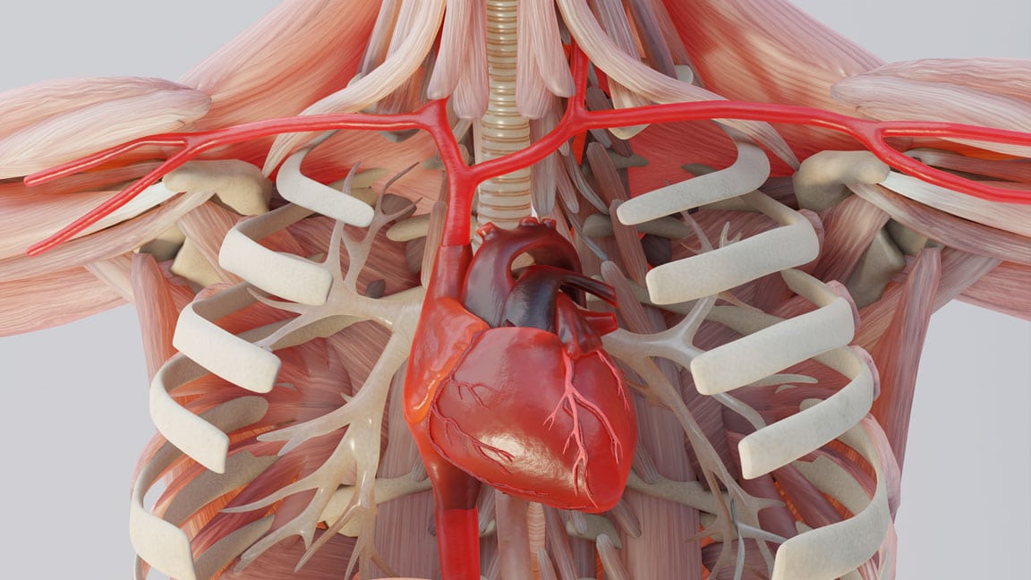

TBI cases don't lose because the injury isn't real. They lose because the jury can't see it. We fix that.

Diffuse Axonal Injury & Axonal Shearing
When the CT Scan Looks Normal but Your Client's Life Is Destroyed — This Is Why.
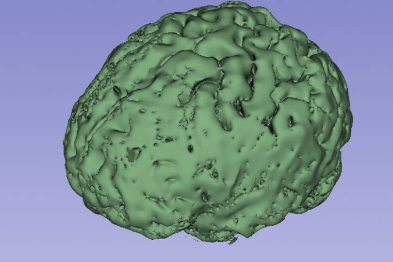
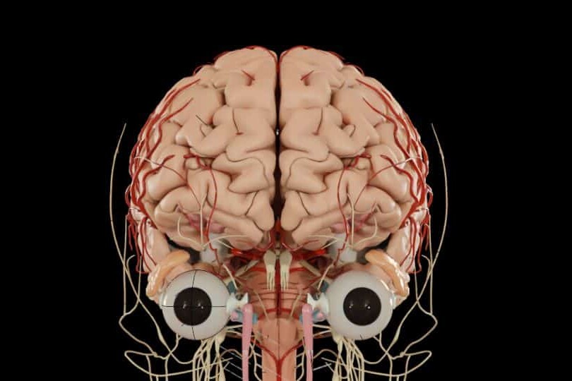
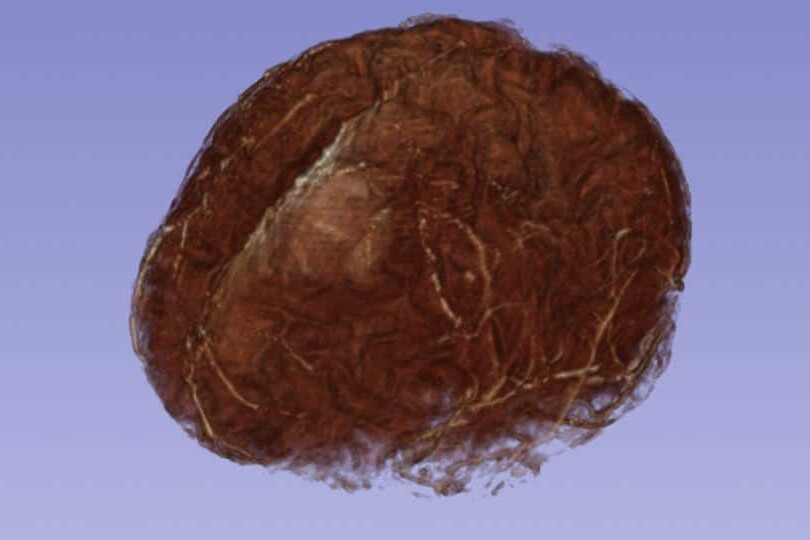
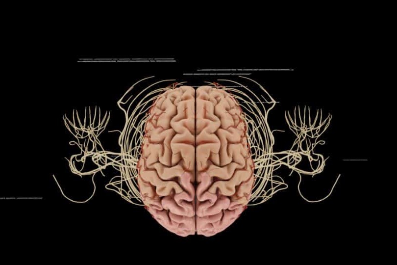
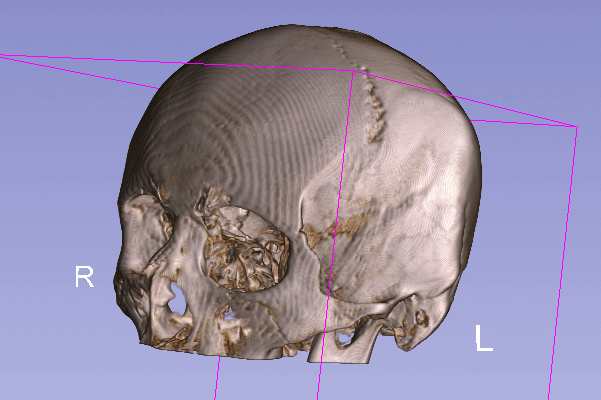
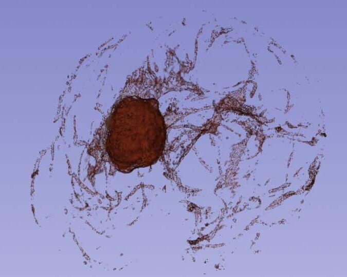
Diffuse axonal injury is the most common and most misunderstood TBI mechanism. The damage is microscopic — scattered across white matter tracts throughout the brain — and invisible on standard CT imaging. Juries see a normal-looking scan and hear an expert describe catastrophic deficits. Without a visual bridge, the gap is insurmountable. We build that bridge. Forensic Reconstruction We animate the mechanics of axonal shearing — the rapid rotational acceleration that stretches and tears microscopic nerve fibers across the corpus callosum, brainstem, and white matter tracts. The physical mechanism of the injury becomes visible, not just described. The Invisible Trauma Using DTI tractography data from your client's own imaging, we render the specific white matter pathways that are disrupted — so the jury sees exactly which neural connections were severed and understands why cognitive, emotional, and behavioral deficits follow even when gross imaging appears unremarkable. DTI Tractography Visualization Diffusion Tensor Imaging data is converted into a 3D rendering of your client's white matter fiber tracts — healthy pathways shown alongside damaged ones. The structural basis for every cognitive deficit your expert has identified becomes a visible, patient-specific exhibit.
Coup-Contrecoup & Kinetic Impact Analysis
One Impact. Two Injury Sites. We Show the Jury Both.
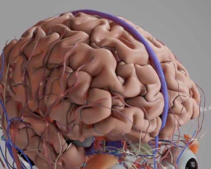
Coup-contrecoup injuries are among the most misunderstood TBI mechanisms. The defense argues your client only struck one side of their head. We show the jury exactly how a single impact produces injury at both the point of contact and the opposite pole — and why the total neurological damage is far greater than a single-site injury. Primary and Secondary Impact We animate the initial coup injury at the point of impact and the countercoup injury on the opposite side of the brain — showing both injury sites simultaneously so the jury understands the full bilateral extent of the trauma, not just the half the defense wants them to see. Kinetic Narrative The physics of the injury are rendered in three dimensions — rotational acceleration, brain displacement within the cranium, and the resulting shear stress across multiple lobes. The jury sees why a single impact produces global neurological compromise across the entire brain, not just at the point of contact. Individual brain lobes are color-coded and labeled — frontal, temporal, parietal, occipital — with injury markers placed at the exact locations documented in your expert's findings. The connection between the physical impact and every reported cognitive, behavioral, and sensory deficit becomes spatially clear.
Intracranial Hemorrhages & Midline Shift
Show the Jury the Bleed Growing — and What It Was Doing to the Brain While It Grew.
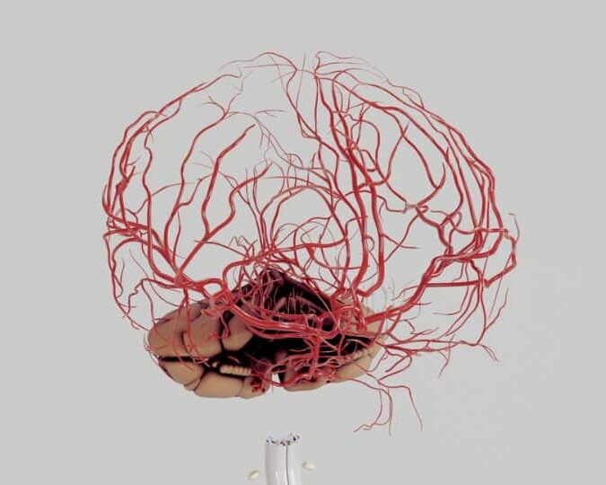
Intracranial hemorrhages are time-critical injuries where delayed diagnosis or intervention directly causes permanent harm or death. We reconstruct the progression of the bleed — from initial rupture to mass effect to herniation — so the jury sees not just that a bleed occurred, but what it did to the surrounding brain tissue and why the timing of intervention mattered. Hemorrhage Mapping Epidural, subdural, subarachnoid, and intracerebral hemorrhages are rendered using Hounsfield Unit-validated data from your client's DICOM imaging — with precise color demarcation of the bleed boundaries, volume, and proximity to critical brain structures. Mass Effect & Herniation The progressive displacement of the midline — derived directly from your client's raw DICOM data — is animated over time to show the physical pressure the expanding hematoma exerted on brain tissue. The jury watches the herniation develop and understands why permanent damage or death was the direct result. Delayed Diagnosis Timeline For cases involving delayed identification or treatment of a hemorrhage, we integrate the bleed progression animation with an interactive timeline showing the window during which intervention should have occurred — and what the imaging showed at each point. The cost of the delay becomes visually undeniable.
Every Animation Is Built From Your Actual Case Records — Not Generic Templates.
Unlike generic animations, our demonstratives are reconstructed from the actual clinical record.
Operative Reports
Step-by-step surgical reconstructions from case documentation.
CT & MRI Imaging
Accurate, DICOM-derived 3D reconstructions of patient anatomy and surgical outcomes.
Expert Testimony
Every animation is developed in collaboration with your retained experts and evolves alongside their opinions.
Medical Literature
Science-backed visualizations grounded in peer-reviewed evidence.
Dedicated Medical Experts For Every Case
Dr. Kevin Ho and his own team of medical experts collaborates directly with your experts for scientifically defensible, strategically aligned medical animations.
Technical Alignment
Every visual element is reviewed against expert reports, deposition testimony, imaging findings, and evolving case facts.
Built on Defensible Science
Animations are built using transparent reconstruction techniques that experts can confidently explain and defend.
Litigation-Focused Collaboration
We help translate highly technical medical concepts into clear visual narratives that resonate with judges, mediators, and juries.
We Stay With Your Case From First Filing to Final Verdict.
Medical negligence cases take years. Expert opinions evolve. New imaging comes in. The case theory shifts. We stay engaged for the full life of the case — refining, updating, and adapting your visual evidence at every stage.
Evolving Forensic Models
As new records, imaging, or expert findings emerge, we update the master 3D models to reflect the most current picture of the injury. Your animation never becomes a liability because it was built too early.
Ongoing Litigation & Deposition Support
We build animations to support long-term litigation — not a single courtroom moment. Demonstratives are adapted through discovery, expert depositions, mediation, and trial, keeping testimony consistent from beginning to end.
The Scientific Credibility Your Opposing Experts Will Recognize
Our work is published by the same institutions cited in medical literature and expert reports. That rigor is what makes our animations defensible — not just persuasive.









Why Top Law Firms Partner with Trial Graphics 360
Unmatched experience and clarity for your case's legal animations.
$100M+
Increased settlement value in high-stakes litigation.
Medically-Trained Animators
Board-certified specialists and former UBC professors ensuring medical accuracy for complex litigation.
Scientifically Published
Harvard, Stanford, and pharma-published expertise giving your case a distinct litigation edge.
Long Term Case Support
End-to-end case support, refining visuals as evidence evolves.
Common Questions About Colorized Diagnostic Films
Standard CT imaging does not detect diffuse axonal injury — the most common catastrophic TBI mechanism. We use DTI tractography data, which measures the integrity of white matter fiber tracts, to render the specific neural pathways your client has lost. The result is a patient-specific 3D exhibit showing structural brain damage that CT simply cannot capture.
At minimum, standard MRI or CT DICOM files. For diffuse axonal injury cases, DTI tractography data produces the most powerful exhibits. We work with whatever imaging is available and can advise on whether additional imaging — such as DTI sequences — should be obtained before expert depositions.
Yes. Our methodology is fully documented — including the specific DTI fractional anisotropy thresholds and Hounsfield Unit values used in each reconstruction. Dr. Ho can provide a complete methodology report and your expert can defend every frame from the stand against opposing expert challenge.
Yes — and TBI animations often produce the most dramatic results at mediation. When an insurance adjuster or defense counsel sees a patient-specific DTI rendering of white matter tract destruction alongside a photograph of your client before the injury, the settlement conversation changes immediately.
Most TBI animations are completed in 4–8 weeks depending on the complexity of the neurological findings and the volume of imaging data. Rush delivery is available — send us your mediation or trial date and we will confirm availability the same day.