


We don't use generic device models. Every reconstruction is built from the manufacturer's actual CAD specifications overlaid on your client's DICOM anatomy — so the jury sees this device failing in this body.


Mechanical Failure & Device Fracture
Show the Jury Exactly Where the Device Broke, Why It Broke, and What the Manufacturer's Own Specifications Say Should Have Prevented It.
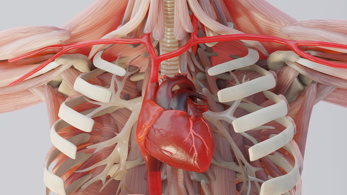
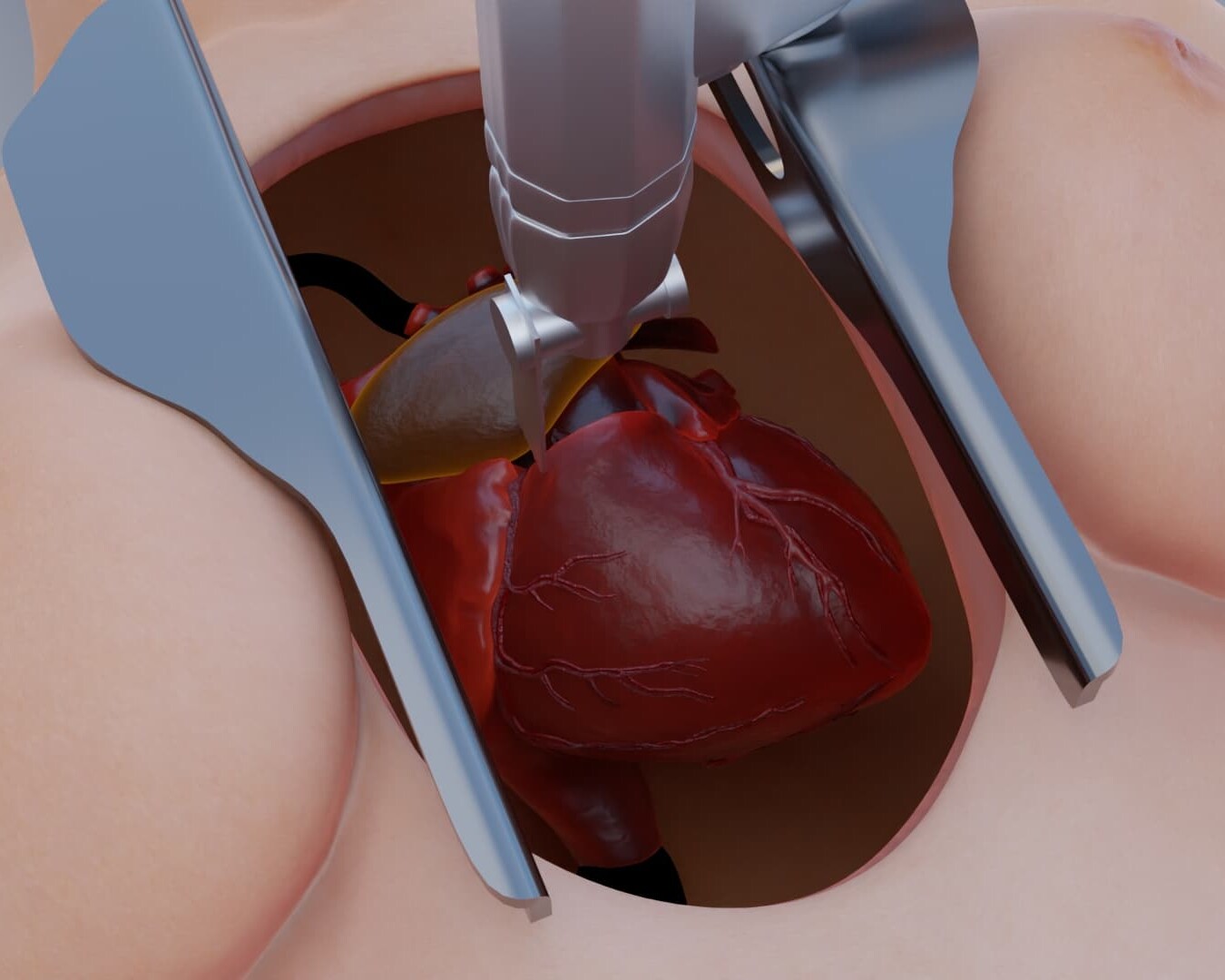
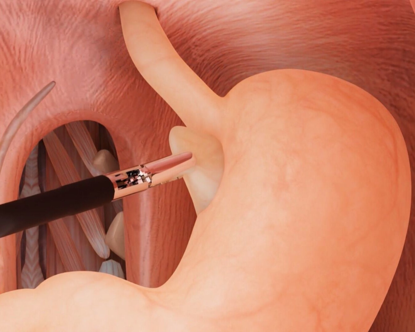
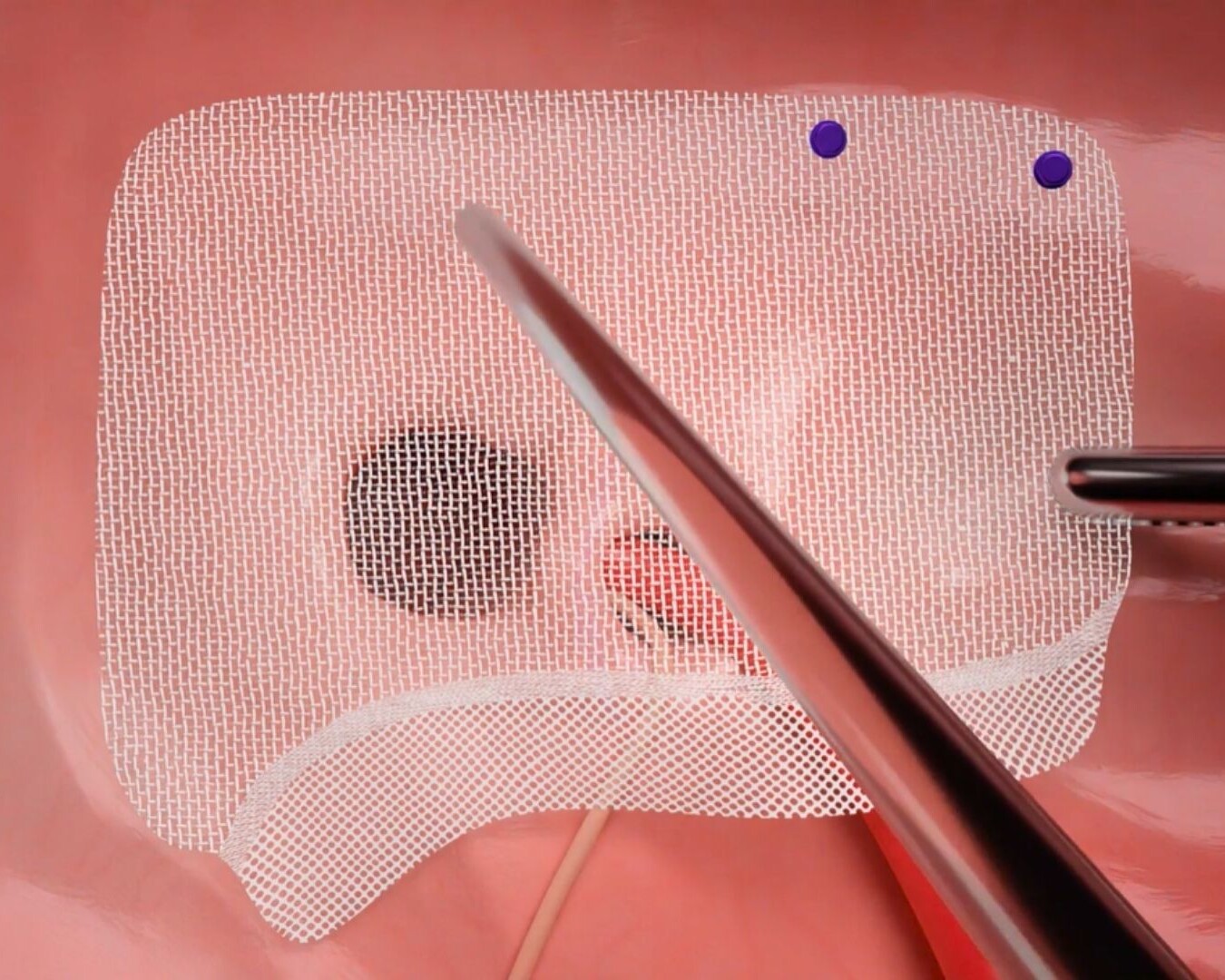
Material fatigue, stress fracture, and structural collapse are engineering concepts that are invisible to a lay jury without a visual model. We build that model — taking the manufacturer's CAD specifications, overlaying them on your client's post-operative DICOM anatomy, and animating the progression from implantation to mechanical failure so the jury sees the failure mode as a physical event, not an abstract engineering opinion. Mechanical Failure & Fracture We animate the specific failure mode — fatigue fracture, stress shielding, cold welding, corrosion, or structural collapse — using the manufacturer's own design specifications and your client's post-operative imaging. Hip stems, spinal screws, rods, cardiac leads, and joint surfaces are all reconstructed with the precision the manufacturer's design documents require. Device Migration & Malposition Using your client's serial DICOM imaging, we animate the progression of device migration from its intended implantation site to the location where it caused secondary injury — showing the path of migration, the structures it encountered, and the vascular, organ, or nerve damage it caused along the way. IVC filter migration, mesh contraction, and implant dislodgement are all reconstructed from the imaging record. Toxicology & Particulate Release We visualize the release of metallic debris, polymer particles, or chemical breakdown products from a failed device — and the inflammatory, fibrotic, or necrotic tissue response in the surrounding anatomy. Metallosis in metal-on-metal hip implants, mesh degradation and erosion, and lead insulation breakdown are rendered at the tissue level so the jury understands the biological consequence of the mechanical failure.
CAD & DICOM Integration — Design Intent vs. Clinical Reality
The Manufacturer's Own Design Specifications Become the Evidence — Overlaid on Your Client's Anatomy.
The most powerful exhibit in a product liability case is the manufacturer's own CAD model placed inside your client's body. It shows the jury what the device was designed to do, what the anatomy required for it to function safely, and exactly where the design failed to account for the clinical reality your client presented. We build that exhibit from the manufacturer's actual design files and your client's actual imaging. CAD & DICOM Integration We overlay the manufacturer's Computer-Aided Design specifications directly onto your client's DICOM anatomy — showing the exact fit, the tolerance gaps, the stress concentration points, and the anatomical variables the design failed to accommodate. The gap between design intent and clinical reality becomes a visual argument, not just an expert opinion. Dynamic Failure Simulation We animate the device under real physiological loading conditions — walking, bending, arterial pulse, respiratory movement, weight bearing — showing how everyday forces your client's body exerted on the device contributed to its failure over time. The jury understands why the failure was foreseeable, not a freak event, and why the manufacturer's warnings were inadequate. Design Defect vs. Manufacturing Defect Side-by-side animations showing the intended design specification alongside the as-manufactured device and the clinical implantation result — isolating whether the failure was a product of the underlying design, a manufacturing deviation, or the interaction between the device and the specific anatomy it was implanted into. Each theory of liability gets its own visual argument.
Device-Specific Reconstructions
Every Device Type Has a Different Failure Mode. We Know Them All.
Product liability cases are won by attorneys who understand the specific failure mechanism of the specific device at issue. We have built forensic reconstructions across every major implant and device category — and we bring that device-specific clinical and engineering knowledge to every case we take. Orthopedic Implants Hip stems, acetabular components, knee implants, spinal hardware, and bone anchors — reconstructed from manufacturer CAD files and post-operative DICOM imaging. Metal-on-metal corrosion, polyethylene wear, stress fracture, and hardware migration are all rendered with the precision your metallurgical and orthopedic engineering experts require. Cardiovascular & Implantable Devices IVC filters, cardiac leads, vascular grafts, stents, and pacemaker components — animated from implantation through failure. Lead fracture, filter strut perforation, stent migration, and graft dehiscence are reconstructed using your client's imaging and the manufacturer's specifications so the failure mode is visually undeniable. Hernia Mesh, Surgical Implants & Drug Delivery Devices Mesh contraction, erosion, and migration into adjacent viscera — reconstructed from post-operative imaging and explant pathology. Surgical stapler misfires, port failures, and drug delivery device malfunctions are animated using the manufacturer's design documents alongside your client's operative and clinical records.
Dedicated Medical Experts For Every Case
Dr. Kevin Ho and his own team of medical experts collaborates directly with your experts for scientifically defensible, strategically aligned medical animations.
Technical Alignment
Every visual element is reviewed against expert reports, deposition testimony, imaging findings, and evolving case facts.
Built on Defensible Science
Animations are built using transparent reconstruction techniques that experts can confidently explain and defend.
Litigation-Focused Collaboration
We help translate highly technical medical concepts into clear visual narratives that resonate with judges, mediators, and juries.
We Stay With Your Case From First Filing to Final Verdict.
Medical negligence cases take years. Expert opinions evolve. New imaging comes in. The case theory shifts. We stay engaged for the full life of the case — refining, updating, and adapting your visual evidence at every stage.
Evolving Forensic Models
As new records, imaging, or expert findings emerge, we update the master 3D models to reflect the most current picture of the injury. Your animation never becomes a liability because it was built too early.
Ongoing Litigation & Deposition Support
We build animations to support long-term litigation — not a single courtroom moment. Demonstratives are adapted through discovery, expert depositions, mediation, and trial, keeping testimony consistent from beginning to end.
The Scientific Credibility Your Opposing Experts Will Recognize
Our work is published by the same institutions cited in medical literature and expert reports. That rigor is what makes our animations defensible — not just persuasive.


Why Top Law Firms Partner with Trial Graphics 360
Unmatched experience and clarity for your case's legal animations.
$100M+
Increased settlement value in high-stakes litigation.
Medically-Trained Animators
Board-certified specialists and former UBC professors ensuring medical accuracy for complex litigation.
Scientifically Published
Harvard, Stanford, and pharma-published expertise giving your case a distinct litigation edge.
Long Term Case Support
End-to-end case support, refining visuals as evidence evolves.
Common Questions About Medical Device Animation
CAD files produce the most precise reconstructions and we request them in discovery whenever possible. However, we can also build accurate device models from manufacturer specifications, implant dimensions from medical literature, explant photographs, and post-operative DICOM imaging. We work with whatever is available and advise early in the case on what documents to request in discovery to support the strongest possible reconstruction.
Explanted devices are often our best evidence. We work from post-explant imaging, pathology reports, metallurgical analysis, and explant photographs to reconstruct the failure mode — and then overlay that reconstruction on the pre-explant DICOM anatomy to show the jury both the device as it failed and the anatomical consequences it left behind.
Yes. We build modular animations that can isolate each theory of liability independently — the design specification against the clinical requirement for one exhibit, the manufacturing deviation against the design specification for another — while maintaining a coherent overall visual narrative that supports both theories simultaneously.
Yes. Our methodology is documented across both disciplines — CAD geometry derivation, DICOM segmentation parameters, physiological loading values, and peer-reviewed materials science and clinical literature citations. Dr. Ho can provide a complete methodology report and your experts can defend the animation from the stand against both engineering and clinical expert challenge.
Most medical device animations are completed in 4–8 weeks depending on the complexity of the failure mechanism and the volume of engineering and clinical data. Rush delivery is available — send us your mediation or trial date and we will confirm availability the same day.