

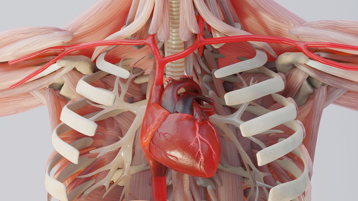

When a medical malpractice or personal injury case goes to trial, the evidence is usually buried inside a stack of MRI images or radiology reports. But in courtrooms, jurors aren’t exactly the radiologists. For them, those MRI slices, CT scan images, and radiology reports were filled with technical terminology. And the process through which jurors can absorb and apply this dense information by breaking down and clarifying the importance of key details is the “DICOM to 3D Model” process.
According to U.S. Legal Support, 37% of law firms utilize specialized, external legal graphics partners for demonstratives, animations, and visual storytelling.
These days, in high-stakes cases where a lot of money is on the line, and medical details matter a great deal, lawyers can’t just hand over those grayscale images to a jury and hope for the best outcomes. Instead, they have to take some medical scans and turn them into something that can tell a story visually. Something that anyone can understand without needing a medical degree. And the best way to do that is by converting the medical imaging data into 3D models and animations. But these models are not just stock animations or generic visuals. These are built from the specific patient data so that the anatomy you are seeing on screen matches the real person.
Learn how you can use these animations to more effectively persuade the jury and accurately represent the medical facts of your case.
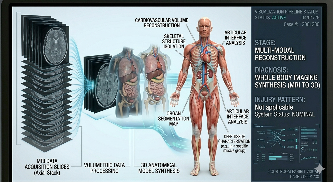
What is the DICOM to 3D Model Process?
So what does it mean, really? When a patient gets an MRI or a CT scan, the machine produces a bunch of raw data. This data is basically like a collection of images that show the slices of the body. If you try to understand things only based on those images, you won’t get anything. The DICOM to 3D model process is a series of steps that takes the raw data and turns it into a 3-dimensional model. Now you can look at that model from any angle, rotate it, and even animate it to show movement or injury.
What is DICOM?
So before we go further, let’s first talk about what DICOM is. DICOM (Digital Imaging and Communication in Medicine) is an international technical standard for medical images and related information. When you get an MRI, the images are saved in this specific format called DICOM, which combines image data (MRI, CT scans, Ultrasounds) with user/patient metadata.
Did You Know?
A CT or MRI scan is not a single image; it is a stack of hundreds of slices, each representing a thin cross-section of anatomy.
Inside a DICOM file, the main thing you’ll find is a series of 2D cross-sectional images. Think of it like a loaf of bread; if you slice it into thin pieces, you’d see a cross-section of everything inside. That’s exactly what an MRI or CT scan does. It takes thin slices of the body and records each one as a separate image.
Beyond just the image, DICOM files also contain a bunch of information about the patient and the scan itself. Things like the patient’s name, the date of the scan, what type of machine was used, how thick the slices are, and technical details about how the scan was performed. Every MRI machine, PET scanner, and CT scanner uses DICOM as its standard. It’s the foundation that all of these systems are built on, which means that no matter where the scan was done or what equipment was used, the data can be accessed and worked with.
Why Raw MRI and CT Data Are Difficult to Use in Court?
When dealing with personal injury cases, it’s important to use medical images to prove what happened. MRIs and CT scans show the injuries, the damage, the surgical site, everything that matters medically. However, the repurpose of an MRI and CT scan is to help doctors figure out what’s wrong with the patient. The images are made for radiologists and surgeons who have years of training in reading them. When you look at an MRI or CT scan, you are seeing a collection of individual images. Each image shows a very thin slice of the body, like a single piece of bread in a loaf. A full scan can include hundreds of these slices stacked together. In each slice, everything is in shades of gray, with different tissues showing up as different levels of brightness. It’s a lot of information, but it’s presented in a format that takes some getting used to. So what’s clear to a doctor is usually confusing for a juror. And when the evidence is confusing, it becomes harder to persuade.
A.) The Core Communication Gap
This creates a fundamental disconnect:
- Radiologists interpret slices
- Attorneys build narratives
- Jurors need clarity
Without a visual transition, this gap leads to:
- Misinterpretation of injury severity
- Confusion about anatomical relationships
- Difficult understanding causation
B. Cognitive Load in Courtrooms
Have you seen one of those courtroom scenes on TV shows? A lot is going on inside the court during a trial session. Witnesses testifying, lawyers asking questions, documents getting exchanged from one party to another. And the jury has to keep track of all that while also paying close attention to the details that matter. When presented with raw images. Jurors have to deal with:
i. Decipher Unfamiliar Visuals: Most people have never looked at an MRI or CT scan before stepping into a jury box/ they don’t know what the different shades of gray mean. They don’t know the difference between bones, tissues, and injury parts.
ii. Decode Medical Terminology: When an expert witness is using medical terms to explain what the images show. Words like “sagittal plane”. “Edema”, “fracture displacement,” or “herniated nucleus pulposus” can be hard to decode with images.
iii. Follow Expert Testimony: White, all this is happening, and images are up on the screen. When the expert is talking, jurors try to look at the images along with them, trying to understand the severity of the situations. It’s a lot for anyone to handle.
The human brain can only process so much information at once. Interpreting unfamiliar visuals, decoding complex language, and following technical testimony, it’s all too much.
Did You Know?
Medical imaging was developed primarily for clinical diagnosis—not for explaining conditions to non-medical audiences, which is why translation into visual formats is often necessary in legal settings.
Complexity Behind “Simple” Scans
Beneath those images of patients lies an enormous amount of structured data. Each image is made up of thousands of tiny data points, and when you put all the images together, you have a complete 3D dataset that contains the exact location, size, and shape of every structure inside the body that the scan captured.
Each scan contains:
- Spatial coordinates
- Tissue density values
- Signal intensities
- Anatomical boundaries
What looks like a simple set of pictures can be a detailed map of someone’s anatomy, but those details are just hidden unless you know how to extract them.
The DICOM to 3D Model Process
DICOM to 3D Model Process is a complex way of producing a structured, visual representation of anatomy from raw data/images.
Let us go into some detail as to what this means:
1. Data Acquisition
This is when attorneys or other medical professionals can provide:
* CT Scans (bone detail)
* MRI Scans (soft tissue detail)
* CBCT (For dental/maxillofacial)
* Clinical Notes and Operations Reports
What Makes Data “Usable”?
The way that someone makes data “usable” is determined by:
Quality – depends on things like:
– Resolution
– Slice Thickness
– Imaging Modalities
– All Available Data
2. Pre-Processing
Prior to reconstructing the 3D image, the raw data from the scanner must be cleaned.
This Includes:
– Noise Reduction
– Contrast Adjustment
– Aligning the Slices
Why is This an Important Step?
Even a minor error in the image data can result in:
– Misinterpretation of anatomy
– Incorrect 3D Reconstruction
– Difficulty When Being Cross-Examined
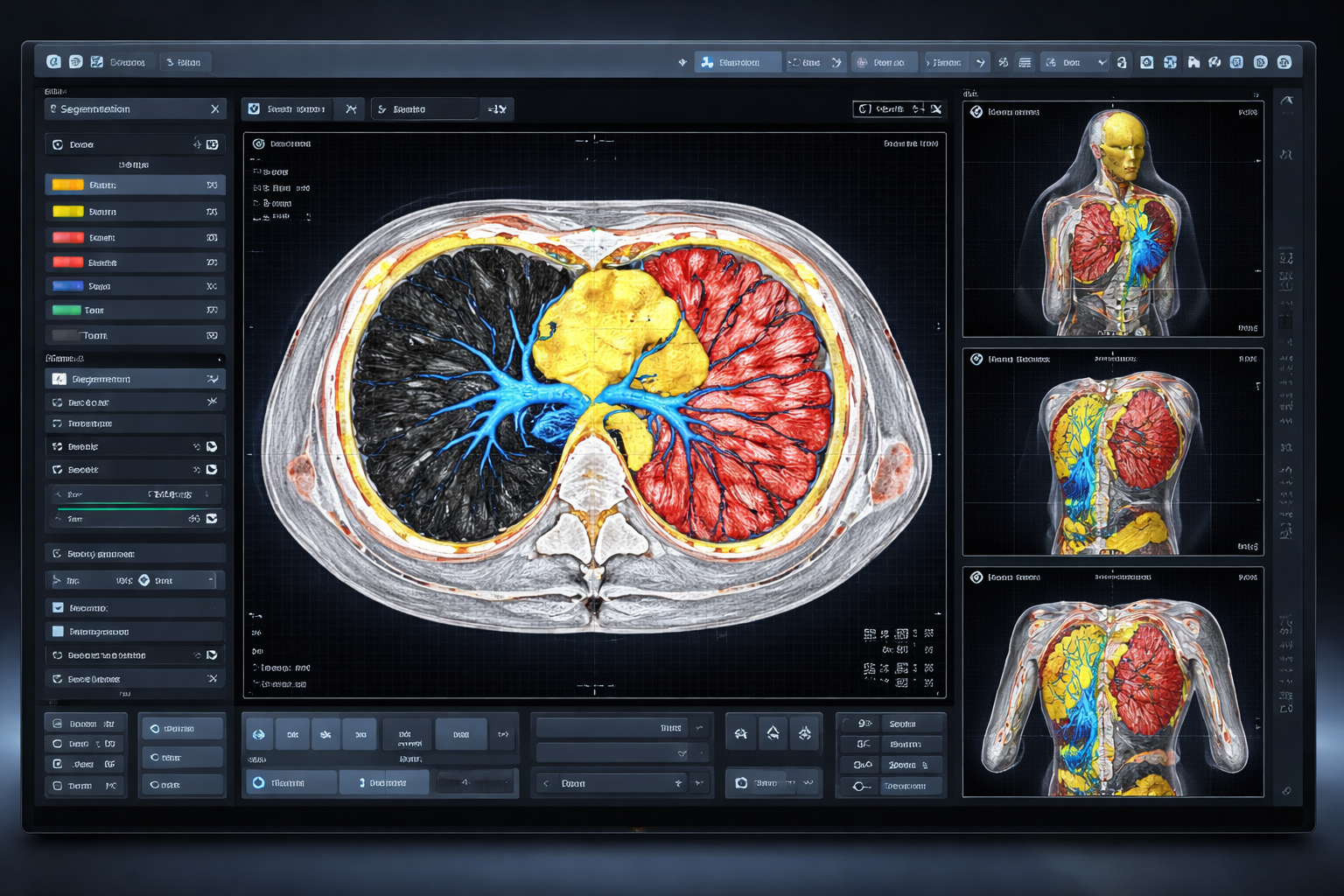
3. Segmentation
This is where all the specialists will identify and isolate the different types of tissue within the anatomy:
What Happens Here?
– Bone
– Nerves
– Blood Vessels
– Soft Tissue
Manual vs Automated Segmentation
| Method | Advantage | Limitation |
| Automated | Fast | Less precise |
| Manual | Highly accurate | Time-intensive |
Why Expertise Is Important
Segmentation involves not only technical procedures, but also interpretation. It incorporates anatomical knowledge, clinical experience, attention to detail, and has associated legal ramifications. Errors in the segmentation of images can change the anatomical representation of the area and/or challenge an argument for causation in court.
4. 3D Reconstruction
3D Models are created by taking the data processed through the segmentation process and rendering it into a 3D image.
The process involves:
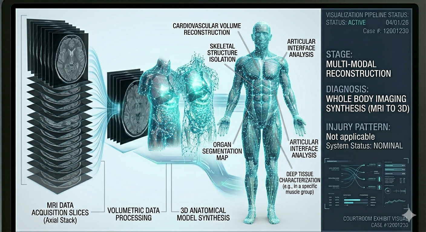 Stacking the slices
- Creating surfaces
- Refining the geometry
Types of Reconstruction
There are several different methods of rendering the 3D model:
Surface Rendering
Volume Rendering
Hybrid models
Why This Step is Powerful
This step is very significant because, for the first time, you can visualize the anatomy spatially and understand it visually.
5. Refinement
Refining the initial model is an essential step to ensure that:
- The anatomy is accurate.
- The surfaces are smooth.
- The surface areas are free of artifacts.
This includes:
- Mesh optimization
- Edge correction
- Validation of structure
Important Notice
The refinement process mustn’t create:
Artistic interpretations
Assumptions not supported by data.
6. Animation
At this stage in the process, the 3D model may be animated and used as a storytelling tool by integrating:
What Animation Adds
- Motion
- Sequence
- Causation
Example Applications
Examples of Uses of Animation Include:
- Showing how a fracture occurred
- Demonstrating how a surgical error was made
- Showing how blood flow through the body changed as a result of an injury
Why Does This Matter?
Animation helps jurors understand:
- Sequence of events
- Timing of the events
- Relationship between cause and effect
7. Validation for Clinical and Legal Purposes
Validation of an animation is required before its use in court.
- Medical Experts Review
- Alignment with testimony
- Consistency with evidence
All animations used in court must first undergo medical reviews by medical experts for validation purposes.
Testimony Support:
All animations used in court must support the testimony of any witness(es) and must not contradict the evidence (research, clinical data) presented.
The Animation Must also Consider:
– Expert opinion
– Avoid speculation
– Neutrality in presentation
The Importance of Transparency in Litigation
Transparency is one of the most significant yet least recognized parts of the DICOM to 3D model process.
Importance of Transparency
Attorneys have a right to know:
– How the model was created
– What data was used
– Who the validators are
Value of Transparency for Attorneys
Transparency provides attorneys with a better opportunity to establish:
– The admissibility of the evidence presented in court
– Credibility of the evidence presented in court
– Persuasiveness of the evidence presented in court below visualization
Establishing a Narrative Beyond Visualization
An effective animation is not only technically accurate; it is also textually effective (narratively).
It Should Answer
Examples of Questions That Should Be Addressed in the Animation:
– What happened?
– How did it happen?
– Why does it matter?
Narrative Flow Example
Normal Anatomical Flow Sequence:
– Normal anatomy (nociceptors)
– Incident (injury/surgery)
– Resultant injury/illness
– Long-term effects
Common Pitfalls Attorneys Should Be Aware Of
As with any graphic evidence used in court, even the best animations can be flawed and therefore ineffective if not utilized properly.
Risks
– Over-simplification of the event depicted in the graphic
– Over-exaggerative presentation of the event depicted in the graphic
– Lack of expert support for the original event being depicted
– Misalignment of the animation with the witness’s testimony
Best Practice for Using Animations in Court
Before using an animation at trial, attorneys must be able to testify as to:
– Evidence foundation for the animation
– Agree with the expert’s opinion of the incident represented in the animation
– Provide a clear explanation of the animation to the jury while at trial
When is the Model Conversion Process Most Effective?
The process of converting DICOM to 3D models is highly effective when the incident being litigated involves:
– Complex anatomy
– Internal organs injuries
– Multiplane imaging
Disputed causation
High-stakes litigation
High Impact Scenarios
Traumatic brain injury
Surgical malpractice
Birth injury cases
Orthopaedic trauma
Future of Medical-Legal Visualization
The way lawyers deal with evidence in court is not the same as it was ten or twenty years ago. Instead, the industry is moving in a few specific directions that are worth paying attention to.
Visual first evidence
Interactive presentations
Data-driven storytelling
Emerging Trends
Beyond the big changes, there are some specific technologies that are starting to show up in courtrooms more often.
Real-time 3D interactions in court
AI-assisted segmentation
Virtual reality demonstrations
Final Thoughts
When you think about what happens when a medical scan becomes an animation, we can only think of the technical steps, like software, segmentation, and rendering. But that misses the bigger point. What is really happening is that the raw data doesn’t mean much to most people, but it’s the real deal. That raw data is being turned into something that can be read by anyone who looks at it.
On one side of this, you have a clinical world full of complex images, technical terms, and information that’s scattered across hundreds of individuals’ scan slices. On the other side, you have the legal world, where clarity matters more than anything, and juries need to understand what happened. The DICO to 3D model process is what connects these two worlds. It takes all those fragmented pieces of imaging data and weaves them into a single coherent story that’s based on real evidence and presented in a visual format that anyone can follow.
It’s not enough to just know that medical animations exist and that they can be useful in court. Lawyers need to understand how these visuals are made, where the data comes from, and what the limitations are. This becomes an even more necessary part of practicing law in cases that involve medical evidence.
Make your medical evidence easier to understand and harder to challenge. Leverage scientifically grounded 3D animations that align with expert testimony with “Trial Graphics 360”.
Frequently Asked Questions (FAQs)
Q.1. What is the DICOM to 3D model process?
The process of DICOM to 3D model is a conversion of medical images, which may include MRI scans or CT scans, into a 3D model.
Q.2. What’s the importance of segmentation
The segmentation process is vital in this case, as it separates different tissues in the images provided. Therefore, this process needs to be closely monitored by experts to ensure accuracy in the 3D model, especially in a legal case.
Q.3. Can MRI data be used for 3D animations?
Yes, MRI scans can be used in this process, which is a conversion of images from a DICOM to a 3D model. This helps in creating animations, which explain injuries or medical conditions.
Q.4. Is the DICOM to 3D model process accurate?
This process can be very accurate, provided it is carried out in a correct manner with quality images.
Q.5. How is the process used in legal cases?
The process of DICOM to 3D model is used in a legal case to explain certain medical conditions or injuries, which helps in creating visuals for a jury to understand complex medical issues in a court of law.