01
Clinical Oversight by Dr. Kevin Ho
Every project is directed by Dr. Kevin Ho, whose background provides a unique "triad" of credibility that is unmatched in the litigation support industry:
Academic Authority
Former Clinical Assistant Professor at the University of British Columbia (UBC) and collaborator with top-tier institutions including Stanford University.
Scientific Validation
A published researcher whose visualizations have been featured in medical research for Harvard and Sanofi.
Clinical Practice
An active Practicing Dentist and Practice Owner, providing real-world insight into surgical standards, patient care, and the "Standard of Care."
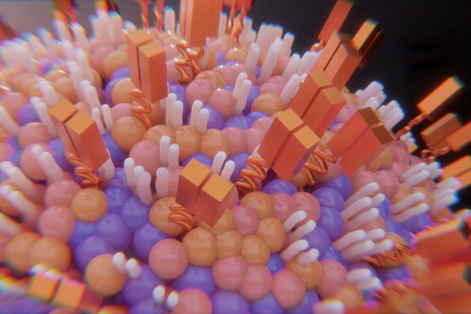
Published Accurate Medical Visualisations in Collaboration with Harvard University, Stanford University, Sanofi and Many More


02
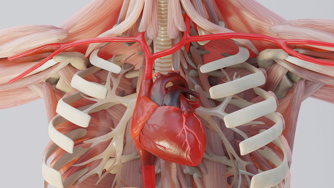
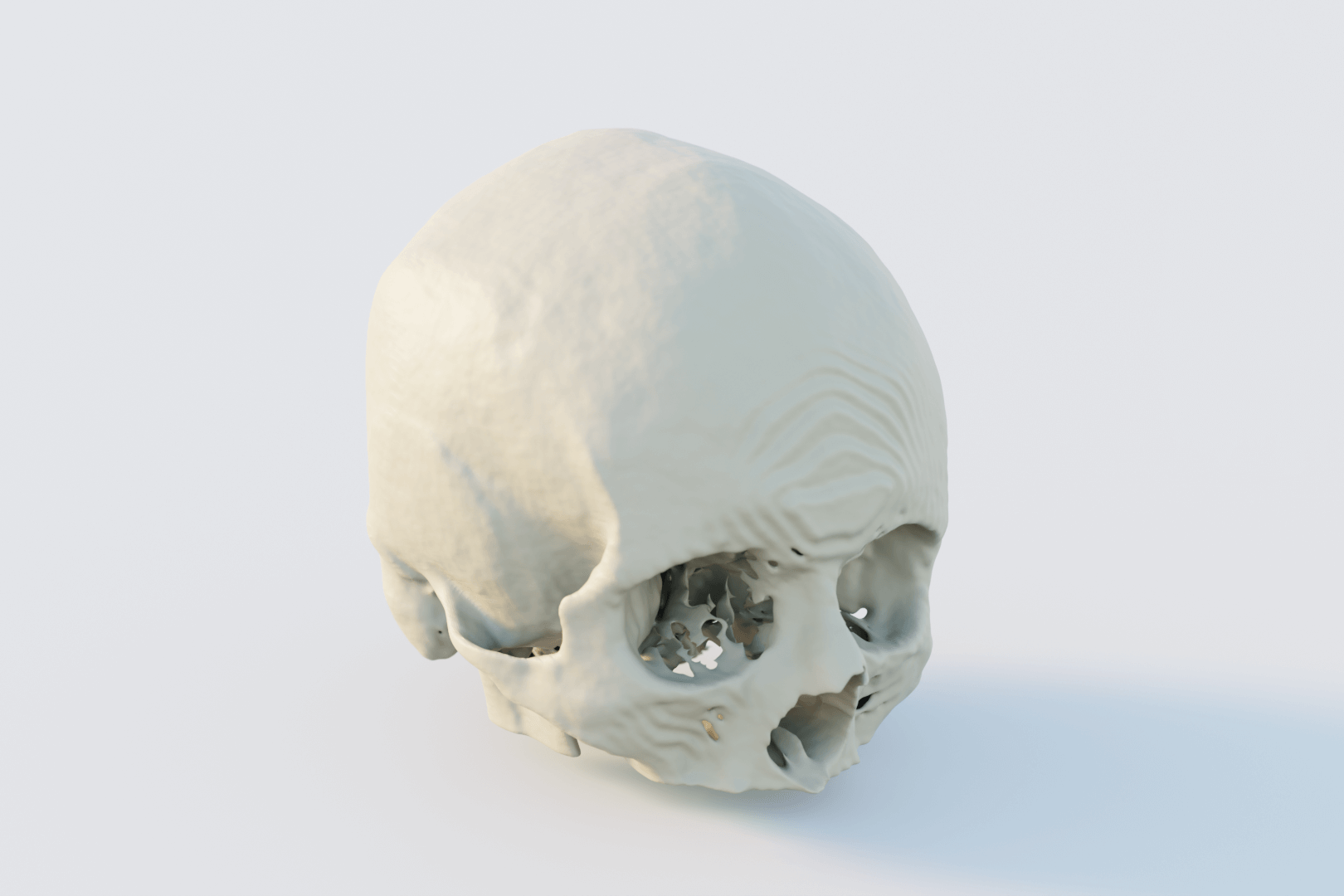
Data Acquisition & DICOM Segmentation
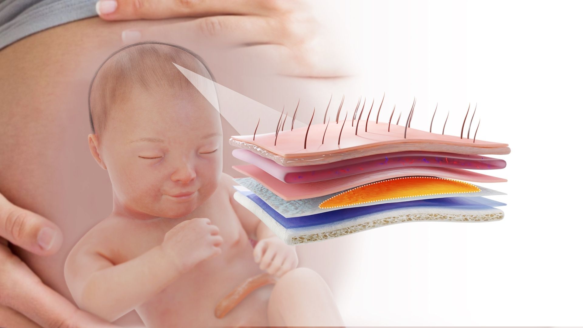
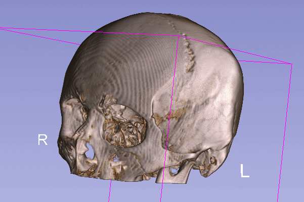
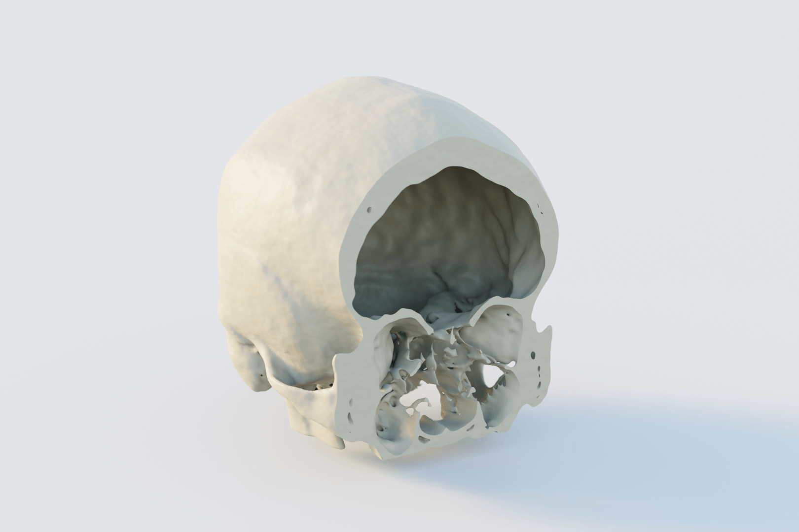
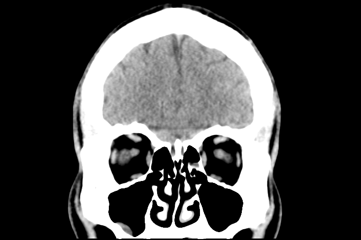
The foundation of every exhibit is the plaintiff’s actual medical imaging and notes. We utilize raw DICOM data (CT, MRI, CBCT) where possible, to ensure the reconstruction is a 1:1 match to the individual’s unique anatomy. We can also utilise clinical notes to reconstruct medical events.
Hounsfield Unit (HU) Thresholding
We apply specific density thresholds to mathematically isolate different tissue types. By setting exact HU ranges, we can separate bone from soft tissue with forensic certainty, removing "artistic license" from the reconstruction.
Segmentation Rigor
Our clinical experts perform manual segmentation to define complex fractures, nerve pathways, or tumor margins. This ensures the final 3D model is a "fair and accurate representation" of the pathology.
Threshold Validation
We provide the technical data-threshold values used in our process, allowing your medical experts to confidently verify the accuracy of the exhibit during deposition or trial.
Accepted Formats: Raw DICOM, CBCT, MRI (T1/T2/DTI), CT.Security: HIPAA-Compliant, forensic-grade encrypted storage.Validation: Hounsfield Unit (HU) thresholding for tissue differentiation.
03
Proven Results & Strategic Impact
Our methodology is designed to do more than just look impressive; it is designed to win. We have a proven track record of helping legal teams maximize the value of their cases.
High-Stakes Success
Our animations have been instrumental in helping legal teams secure landmark results, including a $4.2M settlement for birth injury and numerous other six and seven-figure outcomes.
Admissibility Engineering
Our process is designed to satisfy Daubert/Frye standards and the Federal Rules of Evidence, ensuring your most persuasive evidence actually reaches the jury.
Mechanisms of Injury
We specialize in showing the "invisible" injury—from axonal shearing in the brain to nerve impingement in the jaw—using the same high-end visual standards trusted by global leaders in medicine.
Trusted by the World’s Leading Medical & Academic Institutions.


Visual & Medical Accuracy
Bridging the gap between complex medical data and jury comprehension through precision-engineered 3D visuals.
✳︎ Founded by scientifically published medical illustrators and legal strategy experts
$100M+
Total increase in settlement value for our clients across high-stakes litigation.
15Years
Years of experience in anatomical modeling and courtroom-admissible evidence.
Got some questions?We have answers
Admissibility is built into our foundation. By using Hounsfield Unit (HU) thresholding on the patient’s actual DICOM data, we eliminate “artistic license.” Because every project is overseen by Dr. Kevin Ho (Former UBC Clinical Faculty), we provide a clinical foundation that aligns with the radiology reports, making the exhibit a scientifically-validated representation of the injury.
Yes. Our methodology is based on repeatable, peer-reviewed scientific processes. Because Dr. Ho is a published researcher (Harvard/Sanofi) and a practicing clinician, we can provide the underlying data-threshold values and segmentation protocols used. This transparency allows your medical experts to testify to the accuracy of the model with total confidence.
We provide Strategic Continuity. Unlike “boutique” animation shops, we function as a long-term litigation partner. We maintain the master forensic files throughout the multi-year lifecycle of the case. As new imaging or expert depositions emerge, we update the models to ensure your visual strategy remains current from initial filing to final verdict.
Can you visualize simultaneous liability, such as a medical device failure and surgical malpractice?
This is our specialty. We often manage complex cases involving “finger-pointing” between manufacturers and clinicians. We can concurrently model the mechanical failure of a device and the subsequent clinical response of the surgical team, providing a clear narrative for even the most “tangled” liability webs.
Frequently. Dr. Ho acts as a peer-level bridge between your high-level medical experts and the jury. We speak the technical language of Emeritus Professors and can translate their complex anatomical testimony into clear, persuasive visual assets that are perfectly synchronized with their reports.
We operate with Forensic Neutrality. We are retained by both sides of the bar. Our commitment is to the anatomical data and the clinical record. This reputation for neutrality is often a key factor in having our exhibits admitted without objection during mediation or trial.