


Inguinal Hernia Repair: Visualizing Nerve Entrapment
The Challenge: A patient undergoing a laparoscopic inguinal hernia repair with mesh implantation suffered persistent, debilitating nerve pain post-operatively. Plaintiff's counsel alleged that the surgeon improperly placed the mesh and tacks, leading to entrapment and damage of the ilioinguinal or genitofemoral nerves. The defense contended the pain was a known surgical risk or unrelated.
Our Objective: Uncovering Deviations in Surgical TechniqueVerbal testimony on surgical technique, even with intraoperative photos, often struggles to convey the precise relationship between surgical actions and anatomical injury. Trial Graphics 360 was retained to forensically reconstruct the laparoscopic repair, demonstrating how specific surgical actions deviated from the standard of care and caused nerve entrapment.


The Forensic Solution: Reconstructing the Intraoperative Environment
Using the surgeon’s operative report, post-operative MRI/CT scans (DICOM data), and anatomical guidelines for hernia repair, we built a 3D reconstruction of the inguinal region, the balloon dissection, and the mesh placement.
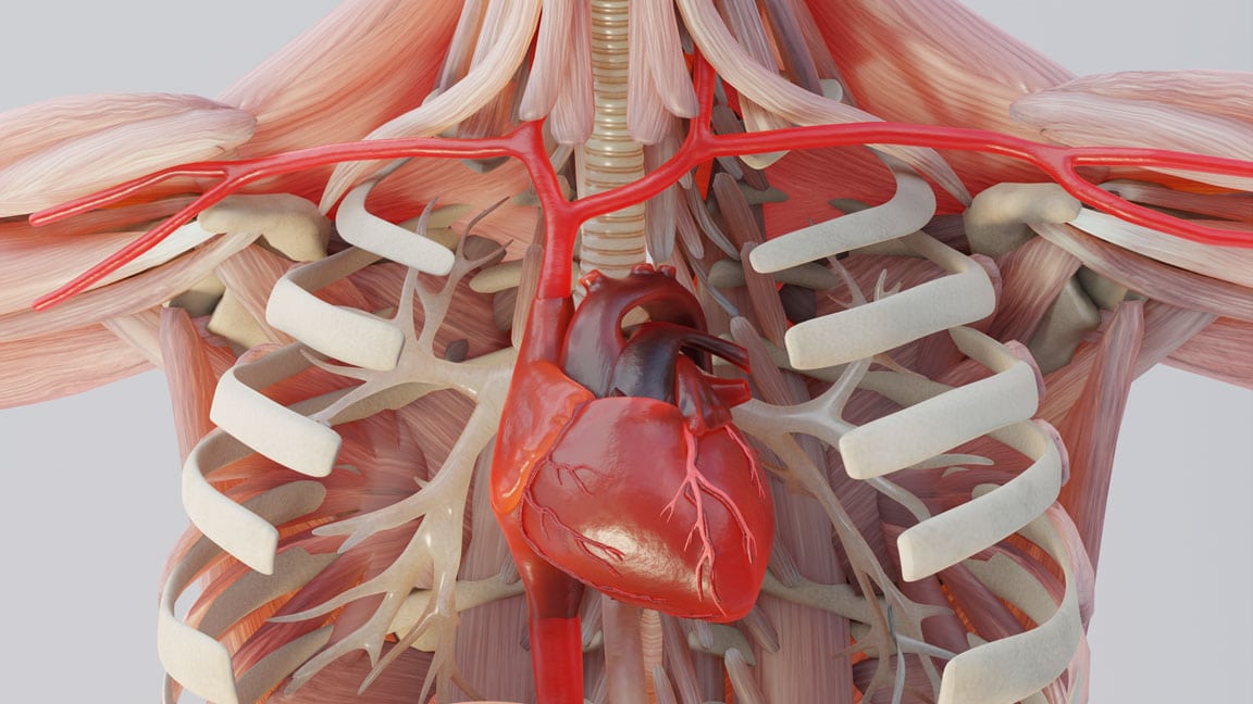
Anatomical Context & Dissection
The animation began by showing the precise pre-operative anatomy, followed by the balloon dissection to create the retroperitoneal space. This established the foundational anatomy and the space available for mesh placement.
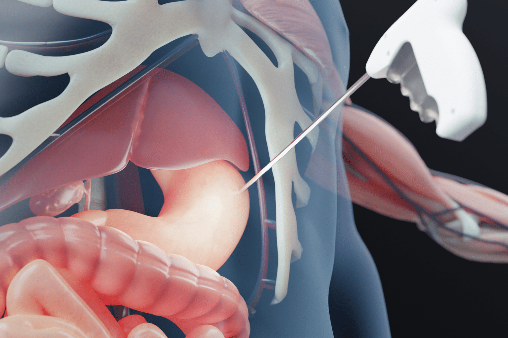
Mesh Placement & Fixation
We meticulously reconstructed the placement of the surgical mesh and the firing of the fixation tacks. The animation highlighted the critical zones where major nerves (ilioinguinal, genitofemoral) traverse the surgical field.
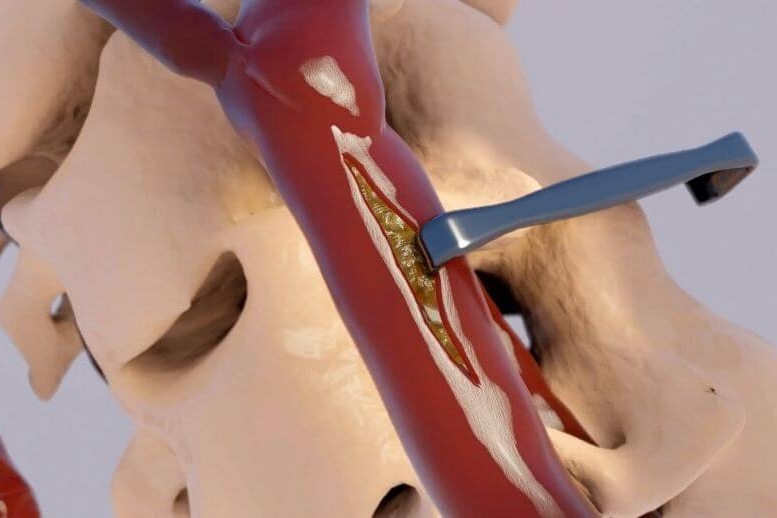
Nerve Entrapment Visualization
By superimposing the reconstructed mesh and tack positions onto the patient's individual nerve pathways (derived from high-resolution imaging), the animation precisely demonstrated how one or more tacks were placed directly onto or in close proximity to the nerve, or how the mesh itself was tensioned incorrectly, leading to entrapment.
Lead Counsel Testimonial
"Dr. Ho's reconstruction of the hernia repair was phenomenal. It allowed us to show, definitively, how the nerve was damaged. The animation clearly depicted the precise placement of the mesh and tacks in relation to the nerve, making our case for negligence undeniable. The defense had no credible counter-argument."
Lead Defense Partner, [Firm Name Redacted]
The Strategic OutcomeResult: Favorable Pre-Trial Settlement for Plaintiff
The forensic animation became an indispensable tool during expert depositions and mediation. Its visual clarity transformed a complex anatomical argument into an undeniable demonstration of surgical error.
Expert Adoption
The plaintiff's surgical expert utilized the animation to precisely illustrate how the nerve was entrapped, providing clear, visual support for their testimony regarding the deviation from standard surgical technique.
Irrefutable Causation
The 3D reconstruction visually established a direct causal link between the tack placement (or mesh tension) and the patient's debilitating post-operative nerve pain.
Increased Settlement Leverage
Faced with the compelling visual evidence, the defense recognized the significant liability exposure, leading to a substantial pre-trial settlement that addressed the patient's chronic pain and long-term care needs.