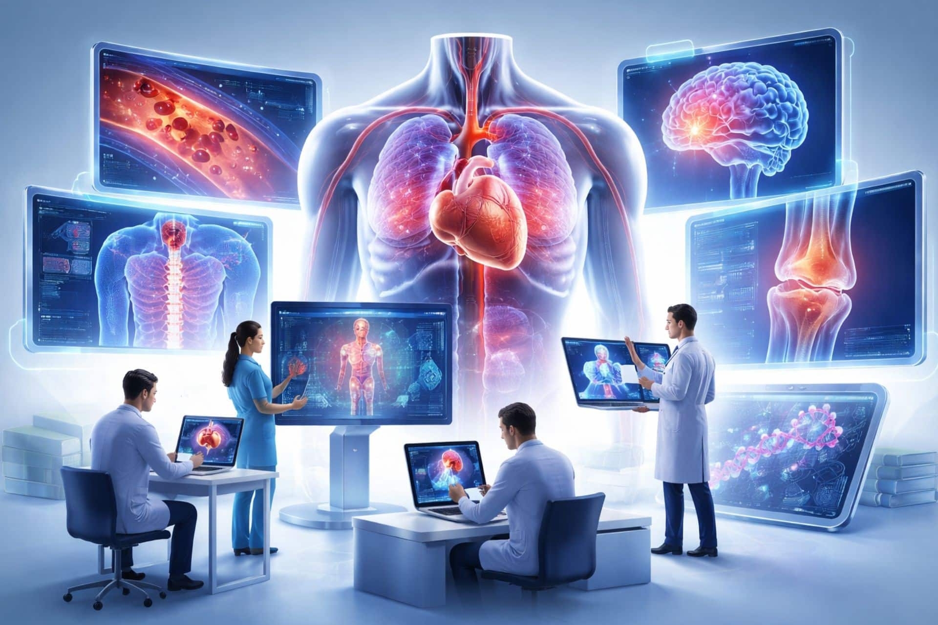
Most trial attorneys are used to receiving a packet of JPEGs or a printed film from a radiology department. They see a 2D gray image and assume they have the “whole picture.”
This is a strategic mistake.
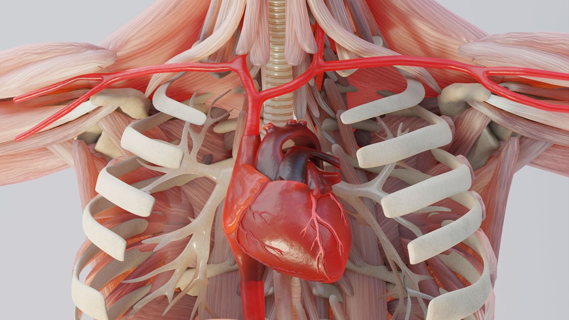
Standard radiology slices are 2D snapshots of 3D problems. If a fracture is non-displaced or a nerve impingement occurs at a specific angle, a 2D slice might miss it entirely. This is why at Trial Graphics 360, we insist on working with the raw DICOM (Digital Imaging and Communications in Medicine) data.\
The Power of 3D Volume Rendering
When we take the raw DICOM data, we can perform what is known as Volume Rendering. This allows us to:
Segment the Anatomy: We can digitally “remove” obstructing bone to see hidden fractures.
Rotate 360 Degrees: We can view the pathology from angles a standard radiologist never considered.
Visualize Impingement: We can show the jury exactly how a herniated disc is physically pinching a nerve root.
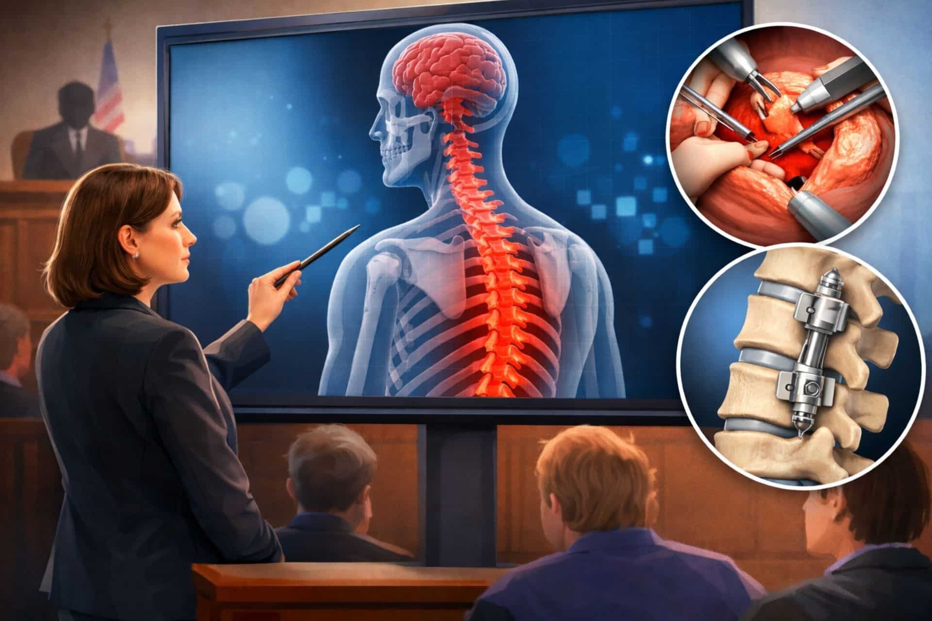
A Message to Counsel
If you are relying on 2D snapshots, you are letting the defense dictate the visual narrative. By using 3D reconstruction, you provide the jury with a “surgeon’s eye view” of the injury, making the pathology impossible to ignore.